. 2012 Apr;32(8):1496-505.
doi: 10.1128/MCB.06554-11. Epub 2012 Feb 13.

In vitro and in vivo antiangiogenic properties of the serpin protease nexin-1

Affiliations

In vitro and in vivo antiangiogenic properties of the serpin protease nexin-1

Sonia Selbonne et al. Mol Cell Biol. 2012 Apr.

Abstract

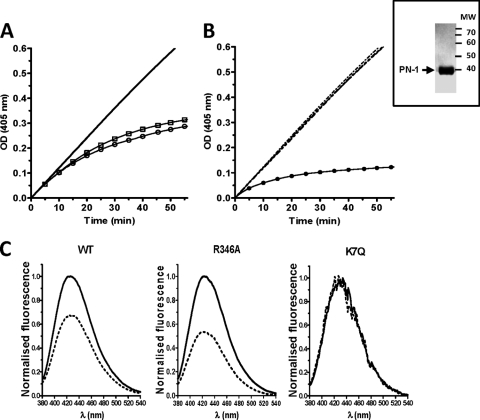
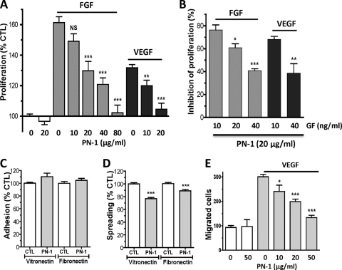
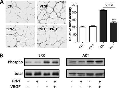
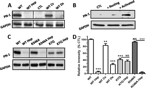
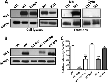
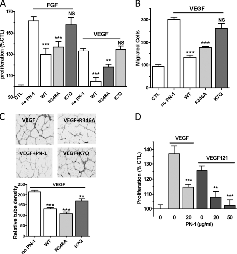
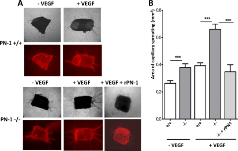
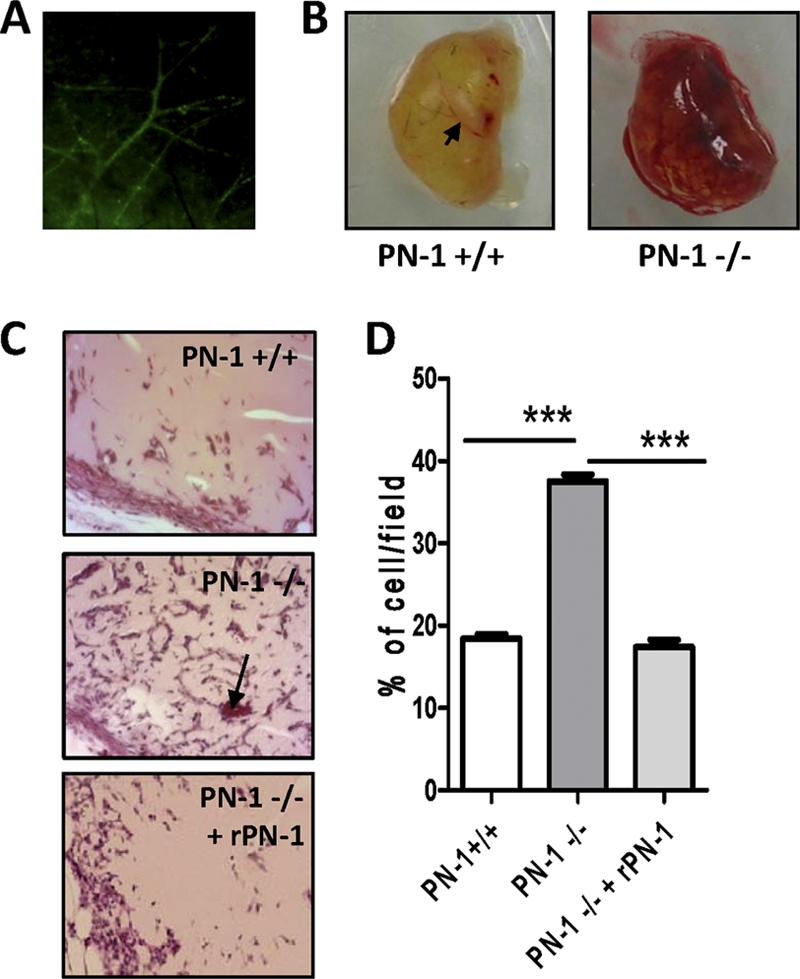
The serpin protease nexin-1 (PN-1) is expressed by vascular cells and secreted by platelets upon activation, and it is known to interact with several modulators of angiogenesis, such as proteases, matrix proteins, and glycosaminoglycans. We therefore investigated the impact of PN-1 on endothelial cell angiogenic responses in vitro and ex vivo and in vivo in PN-1-deficient mice. We found that PN-1 is antiangiogenic in vitro: it inhibited vascular endothelial growth factor (VEGF)-induced endothelial cell responses, including proliferation, migration, and capillary tube formation, and decreased cell spreading on vitronectin. These effects do not require the antiprotease activity of PN-1 but involve PN-1 binding to glycosaminoglycans. In addition, our results indicated that PN-1 does not act by blocking VEGF binding to its heparan sulfate proteoglycan coreceptors. The results obtained in vitro were supported ex vivo in PN-1-deficient mice, where the microvascular network sprouting from aortic rings was significantly enhanced. Moreover, in vivo, neovessel formation was promoted in the Matrigel plug assay in PN-1-deficient mice compared to wild-type mice, and these effects were reversed by the addition of recombinant PN-1. Taken together, our results demonstrate that PN-1 has direct antiangiogenic properties and is a yet-unrecognized player in the angiogenic balance.


Figures

Fig 1
Fig 1
Characterization of PN-1 variants. (Inset) SDS-PAGE Coomassie staining of PN-1 after purification. (A and B) Thrombin inhibition by WT and K7Q PN-1 and catalytic effect of heparin. Thrombin and chromogenic substrate concentrations were 100 pM and 300 μM, respectively. Progress curves are shown. (A) Curves obtained with 50 nM WT (○) and K7Q (□) PN-1, used to determine uncatalyzed rate constants; (B) catalytic effect of 5 nM on thrombin inhibition by 2 nM PN-1. Dashed superimposed lines correspond to WT PN-1, K7Q PN-1, and K7Q PN-1 plus heparin. ●, WT PN1 plus heparin. (C) Representative fluorescence spectra of three of the TNS-bound PN-1 variants in the absence (solid lines) or presence (dashed lines) of heparin, determined with 500 nM WT or K7Q PN-1 and 750 nM R346A. Spectra are difference spectra between TNS plus PN-1 with or without heparin and TNS alone.
Fig 2
Fig 2
Effects of PN-1 on angiogenic responses of HUVECs. (A) Concentration-dependent effect of PN-1 on 10 ng/ml FGF- or VEGF-induced HUVEC proliferation (n = 4 to 10). (B) Effect of PN-1 (20 μg/ml) on cell proliferation induced by increasing growth factor (GF) concentrations (n = 4). Results are expressed as the percentage of inhibition of proliferation, calculated from the ratio of the proliferation induced by each growth factor concentration measured in the presence of recombinant PN-1 to that measured in the absence of PN-1. (C and D) HUVEC adhesion (C) and HUVEC spreading on vitronectin and fibronectin (D), in the absence or presence of 20 μg/ml PN-1 (n = 3 to 4). (E) HUVEC migration in the absence or presence of 20 ng/ml VEGF. *, P < 0.05; **, P < 0.01; ***, P < 0.0001 versus VEGF or FGF alone in panels A and E, versus control (CTL) in panels C and D, and versus 10 ng/ml VEGF or FGF in panel B.
Fig 3
Fig 3
Effects of PN-1 on HUVEC responses to VEGF. (A) Capillary tube formation in Matrigel in the presence or absence of 50 ng/ml VEGF and 20 μg/ml PN-1. Results are expressed as the mean tube density relative to the control without VEGF, measured on 3 fields per well from 3 to 4 experiments. ***, P < 0.0001 versus VEGF alone. (B) Akt and Erk phosphorylation. Whole-cell lysates of HUVECs incubated for 10 min in the presence or absence of 10 ng/ml VEGF and/or PN-1 at 20 μg/ml were immunoblotted with specific anti P-ERK and P-Akt antibodies and with antibodies to the whole protein. The results are representative of at least 3 independent experiments.
Fig 4
Fig 4
PN-1 binding to HUVECs at 4°C. (A to C) Cells were incubated at 4°C with recombinant PN-1 variants (1 μg/ml) in the presence or not of heparin (Hep), chondroitin sulfate (CS), or dermatan sulfate (DS) (A and C) or with supernatant of resting or activated platelets (B). Whole-cell lysates were submitted to immunoblotting with a polyclonal anti-PN-1 antibody and an anti-glyceraldehyde-3-phosphate dehydrogenase (GAPDH) antibody as a protein loading control. Results are shown with representative immunoblots from at least 3 independent experiments (A, B, and C). (D) Densitometric quantification, expressed as the mean intensity (PN-1/GAPDH ratio) relative to control (WT binding), determined for each immunoblot. NS, not significant; **, P < 0.01; ***, P < 0.0001 versus WT or the variants without heparin.
Fig 5
Fig 5
PN-1 binding to HUVECs at 37°C. (A) Whole-cell lysates and cell fractions were obtained after a 2-h incubation of PN-1 variants with HUVECs. Mb, membrane; Cyto, cytosolic. Samples were analyzed as described for Fig. 4, and no signal corresponding to PN-1 was detected in the cytosolic fractions. (B) Whole-cell lysates prepared from HUVECs incubated in the presence or not of heparin (Hep) added at the same time (WT Hep) or 2 h after (WT then Hep), WT PN-1, or of RAP were analyzed as described above. Results are shown in representative blots from at least 3 independent experiments (A and B) or expressed as the mean intensity (PN-1/GAPDH ratio) relative to control WT binding, determined for each immunoblot (C). *, P < 0.05; ***, P < 0.0001, versus WT.
Fig 6
Fig 6
Role of PN-1 functional sites on PN-1 HUVEC angiogenic responses. Effects of PN-1 variants (20 μg/ml [A and C] or 50 μg/ml [B]) on FGF- and VEGF-induced HUVEC proliferation (n = 4 to 10) (A), VEGF-induced HUVEC migration (n = 3) (B), and VEGF-induced HUVEC organization in Matrigel (C). The tube density was measured on 3 fields per well from 3 to 4 experiments and averaged relative to the control without PN-1 and VEGF. (D) Effects of WT PN-1 on VEGF 121-induced HUVEC proliferation. **, P < 0.01; ***, P < 0.001, versus VEGF or FGF alone.
Fig 7
Fig 7
Impact of PN-1 deficiency on ex vivo angiogenesis. Thoracic aortic rings from PN-1-deficient (-/-) and WT (+/+) mice were cultured in a collagen gel in the presence or absence of 50 ng/ml VEGF and 20 μg/ml recombinant WT PN-1. The microvascular networks sprouting from the rings were observed by phase-contrast microscopy and after isolectin B4 labeling (in red), and results were quantified by densitometric analysis. Representative micrographs and results are shown for 6 to 20 rings from 3 to 7 mice. ***, P < 0.001 WT versus PN-1-deficient mice in the presence or absence of recombinant PN-1 (rPN-1).
Fig 8
Fig 8
Impact of PN-1 deficiency on in vivo angiogenesis: vessel formation in Matrigel plugs. Control plugs were implanted in 9 to 10 wild-type mice (PN-1 +/+) or PN-1-deficient mice (PN-1 -/-), and plugs supplemented with 20 μg/ml recombinant PN-1 (rPN-1) were implanted in 3 PN-1-deficient mice (PN-1−/− + rPN-1). (A) Representative fluorescence of the vessels following retroorbital injection of FITC-dextran. (B) Representative plugs from PN-1+/+ and PN-1−/− mice. (C) Representative microphotographs of sections of Matrigel plugs stained with hematoxylin-eosin. Magnification, ×200. Arrows indicate erythrocyte-containing neovessels. (D) Quantification of cell infiltration (4 fields/plug). ***, P < 0.001 for WT versus PN-1-deficient mice and for PN-1-deficient mice versus PN-1-deficient mice administered rPN-1.

References

    1. Asanuma K, et al. 2007. Protein C inhibitor inhibits breast cancer cell growth, metastasis and angiogenesis independently of its protease inhibitory activity. Int. J. Cancer 121: 955–965 - PubMed
    1. Baker JB, Low DA, Simmer RL, Cunningham DD. 1980. Protease-nexin: a cellular component that links thrombin and plasminogen activator and mediates their binding to cells. Cell 21: 37–45 - PubMed
    1. Balsara RD, Ploplis VA. 2008. Plasminogen activator inhibitor-1: the double-edged sword in apoptosis. Thromb. Haemost. 100: 1029–1036 - PMC - PubMed
    1. Boulaftali Y, et al. 2010. Anticoagulant and antithrombotic properties of platelet protease nexin-1. Blood 115: 97–106 - PubMed
    1. Boulaftali Y, et al. 2011. Platelet protease nexin-1, a serpin that strongly influences fibrinolysis and thrombolysis. Circulation 123: 1326–1334 - PMC - PubMed

Publication types

MeSH terms

LinkOut - more resources