. 2013 Jun;34(6):1344-56.
doi: 10.1002/hbm.21516. Epub 2012 Feb 14.

Emotional brain rhythms and their impairment in post-traumatic patients

Affiliations

Emotional brain rhythms and their impairment in post-traumatic patients

Jonathan E Cohen et al. Hum Brain Mapp. 2013 Jun.

Abstract

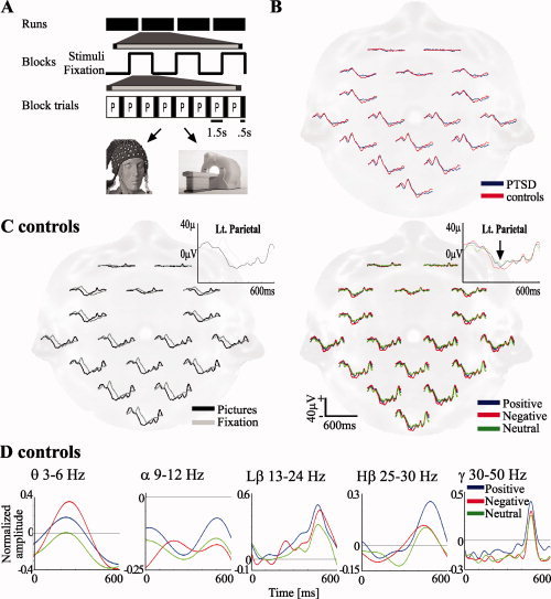
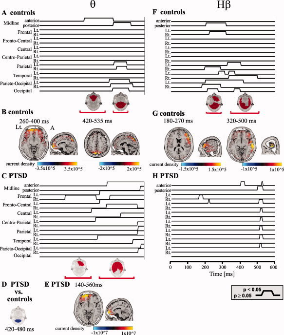
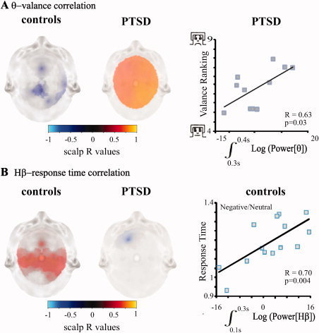
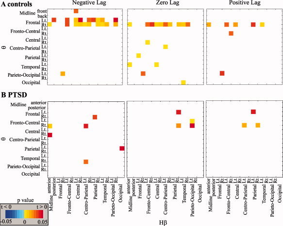
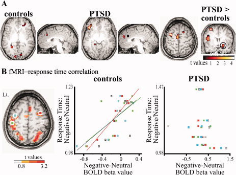
Patients with post-traumatic stress disorder (PTSD) suffer from a failure of cognitive control over emotional distracters. The physiological substrates of cognitive-emotional interactions and their breakdown in disease are, however, unknown. Here, we studied brain activity in PTSD patients and healthy controls in response to emotion-provoking pictures using electroencephalography and functional magnetic resonance imaging (fMRI). We demonstrate that in healthy individuals, emotion-induced frontal theta rhythm modulates activity in the beta rhythm mainly in sensory-motor regions. In contrast, in PTSD patients, beta activity is elevated irrespective of emotion, and is not modulated by frontal theta activity in response to negative emotion. EEG source localization and fMRI findings suggest that theta activity is localized to the prefrontal and anterior cingulate cortices while beta activity is localized to sensory-motor regions. We further found that beta activity in sensory-motor regions is related to the emotion-induced slowing of the motor response in healthy controls while the excess frontal theta activity in PTSD is related to the intensity of negative emotional experience. These findings reveal for the first time the importance of brain electrical oscillations and coherence in emotional top-down modulation and point to specific failure of these mechanisms in PTSD.


Figures

Figure 1
Figure 1
A: Experimental design. Pictures were presented in a block design comprising four runs, each containing three blocks, one of each valence (positive, negative, and neutral) in a pseudo‐randomized order. Each block was preceded by a fixation block. In each block, eight pictures or fixation crosses were presented for 1.5 s, followed by 0.5 s of a black screen each time. B: Visual ERPs in control and PTSD subjects in response to the fixation cross. C: ERPs in control subjects in response to pictures versus fixation (left) and by stimulus emotional valence (right). Insets: enlarged traces. Arrow: negative emotion associated increased negative deflection. D: Averaged (all electrodes) ERS to emotional stimuli at the different frequency bands.
Figure 2
Figure 2
Emotional valence is reflected in spatiotemporal‐frequency alterations. A, C, F, and H: Statistical analysis of the ERSs in the negative versus neutral emotion contrast in the θ (A controls; C PTSD) and Hβ bands (F controls; H PTSD). Statistical significance at a P‐value threshold of 0.05 is plotted for each region and time point. Below are scalp maps of regional statistical significance for representative time windows in which a significant difference was found in the frequency response; red indicates a significant increase, blue a significant decrease at a threshold of P < 0.05. D: Regional statistical significance for comparison of the negative/neutral θ ERS ratio from PTSD to that of controls at 420–480 ms. B, E, and G: Source localization (employing sLORETA) of the signals in response to negative stimuli in the θ (B controls; E PTSD) and Hβ bands (G controls). Lt.: left (for all axial sections), A: anterior (sagittal).
Figure 3
Figure 3
Behavioral correlates of the frequency response. A: Scalp maps of correlation between θ (negative to neutral ratio) and valence ranking of negative stimuli; R values with a P‐value < 0.05 are presented. Time windows with maximal correlation are presented for controls (left, the 400–500 ms post‐stimulus interval) and PTSD (center, the 300–400 ms interval). An example of correlation in the 300–400 ms interval in the right parietal region is shown for PTSD (right; one extreme value was removed from figure, correlation with this value was higher: R = 0.84; P = 0.0006). B: Scalp maps of correlation between the negative to neutral ratios of Hβ activity in the 100–300 ms interval and RTs; R values with a P‐value < 0.05 are presented for controls (left) and PTSD (center). An example of correlation in the 100–300 ms interval in the left central scalp region for controls is shown (right).
Figure 4
Figure 4
Emotion‐induced frontal θ activity leads to widespread Hβ activity in control but not PTSD subjects. A and B: Cross‐correlation analysis between θ and Hβ activity across scalp regions; P‐values for the statistical comparison of response to negative versus neutral emotion for negative, zero, or positive lags of the θ signal in relation to Hβ are shown for controls (A) and PTSD (B).
Figure 5
Figure 5
fMRI analysis points to anatomical regions of emotion‐related activity. A: MRI BOLD signal analysis for the contrast negative‐neutral in control, PTSD and for the contrast PTSD negative‐neutral versus controls negative‐neutral. Encircled is the amygdala. B: Correlation between the difference in individual negative versus neutral mean BOLD activation and ratio of negative/neutral RT in left premotor (blue), left motor (green), right premotor (red), and right motor (gray) regions (left, regions for analysis were extracted from the contrast: all pictures vs. fixation,) presented for controls (center) and PTSD (right). For all fMRI analysis shown in this figure, P < 0.01 uncorrected, cluster size > 5 voxels.

References

    1. Aftanas LI, Varlamov AA, Pavlov SV, Makhnev VP, Reva NV ( 2001): Affective picture processing: Event‐related synchronization within individually defined human theta band is modulated by valence dimension. Neurosci Lett 303: 115–118. - PubMed
    1. Bishop SJ ( 2007): Neurocognitive mechanisms of anxiety: An integrative account. Trends Cogn Sci 11: 307–316. - PubMed
    1. Bishop SJ ( 2008): Neural mechanisms underlying selective attention to threat. Ann N Y Acad Sci 1129: 141–152. - PubMed
    1. Blair KS, Smith BW, Mitchell DG, Morton J, Vythilingam M, Pessoa L, Fridberg D, Zametkin A, Sturman D, Nelson EE, Drevets WC, Pine DS, Martin A, Blair RJ ( 2007): Modulation of emotion by cognition and cognition by emotion. Neuroimage 35: 430–440. - PMC - PubMed
    1. Boashash B ( 1992): Estimating and Interpreting the Instantaneous Frequency of a Signal .1. Fundamentals. Proc IEEE 80: 520–538.

Publication types