. 2013 Jun;34(6):1464-76.
doi: 10.1002/hbm.22003. Epub 2012 Feb 14.

Cortical plasticity is preserved in nondemented older individuals with severe ischemic small vessel disease

Affiliations

Cortical plasticity is preserved in nondemented older individuals with severe ischemic small vessel disease

Jonathan List et al. Hum Brain Mapp. 2013 Jun.

Abstract

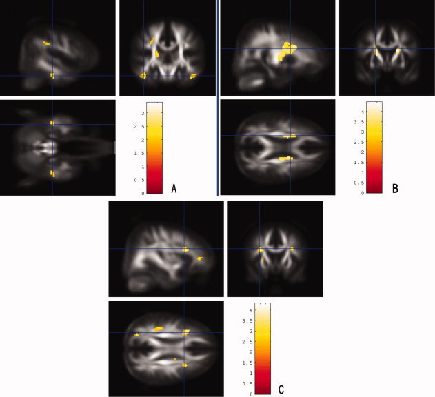
Ischemic small vessel disease (SVD) is a common finding on routine scans in older people, but cognitive sequelae vary considerably. To improve understanding of mechanisms underlying decline or preservation of cognitive function in this condition, we assessed cognition and cortical plasticity in 20 elderly subjects with severe SVD and 20 age-matched controls without SVD, as rated on conventional MRI. Cognitive status was determined with a neuropsychological test battery, cortical plasticity induced with a paired associative stimulation protocol. Microstructural white matter changes were further analyzed for fractional anisotrophy using diffusion tensor imaging. We found that cortical plasticity as well as memory functions were preserved in severe SVD, while executive functions showed trendwise or significant decreases. Within the SVD group, lower white matter integrity in parahippocampal regions and posterior parts of the corpus callosum was associated with larger cortical plasticity, an association not seen for prefrontal white matter tracts. Enhanced cortical plasticity in subjects with lower white matter integrity in memory-relevant areas might thus indicate a compensatory mechanism to counteract memory decline in severe SVD.


Figures

Figure 1
Figure 1
Defined regions of interest (ROI; in red), superimposed on an average FA template of 160 healthy individuals (not shown: ROI of the parietal lobes). [Color figure can be viewed in the online issue, which is available at wileyonlinelibrary.com.]
Figure 2
Figure 2
Mean MEP‐changes (MEAN‐LTP) following PAS in the SVD group and controls. MEAN‐LTP was assessed by the grand average of MEPs at time points T0 to T30, normalized to the baseline MEP. Values > 1 indicate responders to PAS, whereas values ≤ 1 indicate nonresponders to PAS. Although 16 of 20 responded to PAS, only 10 of 20 subjects of the SVD group were responders to PAS.
Figure 3
Figure 3
Rapid‐onset plasticity in patients and controls. Error bars indicate standard error of the mean. No significant differences between both groups could be observed at each time point. * indicate significant changes in MEPs following PAS within a group, as compared to baseline. * = P < 0.05.
Figure 4
Figure 4
Differences in microstructural white matter integrity. SPM “glass brain” representation (left) and statistical FA maps that were superimposed on an averaged FA template (left) showing clusters of decreased FA in the SVD group compared to controls (ANCOVA, modeling age as a covariate; P < 0.001, corrected for multiple comparison; minimum of 25 contiguous voxels). Colored bars represent t‐values; display threshold is set at t‐value > 3.16. FA values of the SVD group were significantly reduced in white matter areas covering widespread parts of the brain, indicating structural white matter changes extending beyond the white matter lesions that showed up on conventional MRI. [Color figure can be viewed in the online issue, which is available at wileyonlinelibrary.com.]
Figure 5
Figure 5
Statistical FA maps that were superimposed on an averaged FA template showing clusters of correlation between MEP changes after PAS and decreased FA values in the SVD group (P < 0.001, minimum of 50 continuous voxels; corrected for multiple comparisons). Colored bars represent t‐values; display threshold is set at t‐value > 3.71. Low FA values particularly in clusters bilaterally in the parahippocampal area, in connecting fibers of the corpus callosum (mainly the posterior part), and parts of the corticospinal tract were associated with greater rapid‐onset cortical plasticity, whereas prefrontal regions did not show this association. [Color figure can be viewed in the online issue, which is available at wileyonlinelibrary.com.]
Figure 6
Figure 6
Brain regions in which a reduced FA was associated with decreased cognitive test results in the SVD group. Statistical FA maps that were superimposed on an averaged FA template. Colored bars represent t‐values; display threshold is set at t‐value > 3.71 (B and C) and > 2.34 (A), respectively. A: FA maps showing clusters of correlation between decreased FA and reduced performance in “working memory” (P < 0.005, > 25 continuous voxels; corrected for multiple comparisons). FA values were lower with decreased test results mainly in bilateral parahippocampal clusters. B: Lower FA values were associated with decreased “verbal memory” test results in frontal parts of the corticospinal tract (P < 0.001, > 50 continuous voxels; corrected for multiple comparisons). C: FA values were lower with decreased “executive function” test results in prefrontal regions and in a cluster in the left parietal region (P < 0.001, > 50 continuous voxels; corrected for multiple comparisons). [Color figure can be viewed in the online issue, which is available at wileyonlinelibrary.com.]

Similar articles

Cited by

References

    1. Arsava EM, Rahman R, Rosand J, Lu J, Smith EE, Rost NS, Singhal AB, Lev MH, Furie KL, Koroshetz WJ, Sorensen AG, Ay H ( 2009): Severity of leukoaraiosis correlates with clinical outcome after ischemic stroke. Neurology 72: 1403–1410. - PMC - PubMed
    1. Aschenbrenner S, Tucha O, Lange K ( 2001): Regensburger Wortflüssigkeitstest (RWT). Göttingen, Bern: Hogrefe‐Verlag GmbH & Co. KG.
    1. Barnes DE, Covinsky KE, Whitmer RA, Kuller LH, Lopez OL, Yaffe K ( 2009): Predicting risk of dementia in older adults: The late‐life dementia risk index. Neurology 73: 173–179. - PMC - PubMed
    1. Battaglia F, Wang HY, Ghilardi MF, Gashi E, Quartarone A, Friedman E, Nixon RA ( 2007): Cortical plasticity in Alzheimer's Disease in humans and rodents. Biol Psychiatry 62: 1405–1412. - PubMed
    1. Bella R, Ferri R, Pennisi M, Cantone M, Lanza G, Malaguarnera G, Spampinato C, Giordano D, Alagona G, Pennisi G ( 2011): Enhanced motor cortex facilitation in patients with vascular cognitive impairment‐no dementia. Neurosci Lett 503: 171–175. - PubMed

Publication types