. 2012 Feb 28;109(9):3359-64.
doi: 10.1073/pnas.1115967109. Epub 2012 Feb 13.

Mechanical stress-activated integrin α5β1 induces opening of connexin 43 hemichannels

Affiliations

Mechanical stress-activated integrin α5β1 induces opening of connexin 43 hemichannels

Nidhi Batra et al. Proc Natl Acad Sci U S A. .

Abstract

The connexin 43 (Cx43) hemichannel (HC) in the mechanosensory osteocytes is a major portal for the release of factors responsible for the anabolic effects of mechanical loading on bone formation and remodeling. However, little is known about how the Cx43 molecule responds to mechanical stimulation leading to the opening of the HC. Here, we demonstrate that integrin α5β1 interacts directly with Cx43 and that this interaction is required for mechanical stimulation-induced opening of the Cx43 HC. Direct mechanical perturbation via magnetic beads or conformational activation of integrin α5β1 leads to the opening of the Cx43 HC, and this role of the integrin is independent of its association with an extracellular fibronectin substrate. PI3K signaling is responsible for the shear stress-induced conformational activation of integrin α5β1 leading to the opening of the HC. These results identify an unconventional function of integrin that acts as a mechanical tether to induce opening of the HC and provide a mechanism connecting the effect of mechanical forces directly to anabolic function of the bone.


Conflict of interest statement

The authors declare no conflict of interest.

Figures

Fig. 1.
Fig. 1.
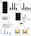
Integrin α5β1 interacts directly with Cx43, and fluid flow enhances the interaction. (A) Colocalization of α5 and Cx43 on the cell surface was detected by dual-immunolabeling of impermeable osteocytic MLO-Y4 cells. (Scale bar, 10 μm.) (B) Cell lysates (lanes 1 and 6) and immunoprecipitates (IP) with antibody to either Cx43 (lanes 2 and 3) or integrin α5 (lanes 4, 5, 7, and 8) were probed for integrin α5 (lanes 1–3), Cx43 (lanes 4 and 5), or integrin β1 (lanes 6–8). (C) The eluted fractions of GST-Cx43CT or GST from beads conjugated with α5 C-terminal or scrambled peptide were immunoblotted using anti-Cx43 (WB, Upper) or were stained with Coomassie Blue (CB, Lower). (D) A concentration series of α5 C-terminal peptide (0.015–2 mM) was injected at 100 μL/min in duplicate over a surface plasmon resonance sensor chip (CM5) to which GST-Cx43CT (specific surface) and GST (reference surface) were covalently attached. (Upper) Double-referenced data were plotted (black traces) and globally fit (red traces) to a 1:1 interaction model using Scrubber 2 software to determine the indicated kinetic and equilibrium constants (Inset). (Lower) Equilibrium analyses were performed by plotting the binding response at equilibrium as a function of α5 concentration and fitting the data to a hyperbolic equation with Scrubber 2 software. (E) Fluid flow increases the interaction. Preloaded cell lysates (Lower) and immunoprecipitates of α5 antibody (Upper) with (FF) or without fluid flow (C) were probed for Cx43 (Left), and band intensity was quantified (Right). P < 0.01, fluid flow (FF) versus control (C).
Fig. 2.
Fig. 2.
Integrin α5 and its interaction with Cx43 are critical for HC opening. (A) MLO-Y4 cells were transfected with α5 siRNA and siRNA from scrambled sequence or transfection reagent (vehicle). ***P < 0.001, α5 siRNA (60 or 90 nM) versus vehicle and scrambled siRNA. (B) Cells were subjected to static (C) or fluid-flow (FF) conditions, and LY dye uptake was quantified. Dye uptake was abolished in α5 siRNA (60 nM)-transfected cells (FF+α5 siRNA). ***P < 0.001, fluid flow versus FF+α5. (C) (Upper) Lysates of MLO-Y4 cells transfected with Cx43CT-GFP or vector (vehicle) were immunoblotted with anti-Cx43CT antibody (Cx43C). (Lower) α5 antibody primarily coimmunoprecipitated Cx43CT-GFP but not endogenous Cx43 in cells overexpressing Cx43CT-GFP. The immunoprecipitates were blotted with antibody to α5, Cx43C, or GFP. The asterisk indicates the heavy chain of IgG. (D) Overexpression of Cx43CT-GFP, but not GFP or vector (vehicle), in MLO-Y4 cells prevented the opening of the HC in response to fluid flow. The number of cells containing GFP taking up Alexa Fluor 350 dye under fluid-flow (FF) or static (C) conditions was quantified. ***P < 0.001, Cx43CT-GFP (static and fluid-flow conditions), GFP (static condition), and vehicle (static condition) versus GFP (fluid-flow condition) and vehicle (fluid-flow condition).
Fig. 3.
Fig. 3.
The binding of FN substrate is not essential for the role of integrin in HC opening by fluid flow. (A) MLO-Y4 cells were cultured on collagen (Col), FN, or polylysine (poly-Lys) matrices and then were subjected to static (C) or fluid-flow (FF) conditions. ***P < 0.001, fluid flow (collagen or FN) versus the corresponding static condition (collagen or FN). (B) Neither FN nor RGD had any effect on the opening of the HC by fluid flow. ***P < 0.001, fluid flow (Ctrl, RGD, or FN) versus the corresponding static condition (Ctrl, RGD, or FN). (C) The number of cells attached and detached in the supernatant was quantified. ***P < 0.001, RGD (supernatant or attached) versus the corresponding static condition (supernatant or attached). (D) Dye uptake was analyzed, and only cells attached to FN beads were quantified. ***P < 0.001, fluid flow (FN-beads or control) versus the corresponding static condition (FN-beads or control). (E) MLO-Y4 cells were cultured on collagen-coated plates for 8 or 48 h and were subjected to static (C) or fluid-flow (FF) conditions. ***P < 0.001, fluid flow for 8 or 48 h versus the static condition for 8 or 48 h. (F) Nonpermeable MLO-Y4 cells cultured on collagen matrix for 8 or 48 h were coimmunolabeled with α5 and FN antibodies. (Scale bar, 50 μm.) (G) MLO-Y4 cells were cultured for 8 h on collagen-coated plates in FN-depleted (−FN) or normal (Norm) medium. ***P < 0.001, fluid flow in medium with or without FN versus the static condition in medium with or without FN.
Fig. 4.
Fig. 4.
Activation of integrin α5β1 by mechanical stimulation is necessary for HC opening, and fluid flow-activated PI3K signaling is involved. (A) MLO-Y4 cells were incubated with coated magnetic beads, followed by the application of a magnetic field. Dye uptake was quantified only in cells attached to magnetic beads. HC opening was increased significantly in cells attached to the beads coated with α5 antibody or FN but not in cells attached to the beads coated with polylysine (Poly-Lys), sheep IgG, or CD44 antibody. ***P < 0.001, FN and α5 antibody versus poly-lysine and CD44 antibody. (Scale bar, 50 μm.) (B) Cells cultured in serum without FN were incubated with mouse IgG or β1-activating antibody (β1 Ab) alone or were pretreated with either carbenoxolone (Carb) or HC-blocking Cx43 (E2) antibody before dye uptake. **P < 0.01, β1 antibody versus all others. (C) Fluid flow stimulates integrin activation. Lysates of fluid flow-treated cells incubated with GST-FNIII9–11 were immunoblotted with anti-GST antibody for detection of bound GST-FNIII9–11 or β-actin. (D) (Upper) Flow-induced HC opening. Compared with the static condition (C), the increased dye uptake in MLO-Y4 cells by fluid flow (FF) was decreased significantly with LY294002 (FF+LY294002). *P < 0.05. (Lower) The interaction between α5 and Cx43 is blocked by LY294002). (E) Fluid flow activates integrin α5β1 through PI3K. MLO-Y4 cells were pretreated for 30 min with the PI3K inhibitors LY294002 (LY, 10 μM) or wortmannin (WM, 100 nM) or were not treated as control (C) before fluid flow for 15 min. Cell lysates were immunoblotted for bound GST-FNIII9–11 or β-actin. Bound GST-FNIII9–11 was normalized with β-actin. **P < 0.01. (F) Schematic diagram of the role of integrin α5β1 in regulating Cx43 HC opening. (Left) In the absence of mechanical loading (static condition), the association between α5β1 and Cx43 exists through their C termini; however, the HCs remain closed. (Right) Upon fluid flow, PI3K signaling is activated, leading to activation of α5β1, promoting conformational change of its extended extracellular domain, and subsequently to the opening of the HC, which allows the passage of small bone anabolic factors, such as prostaglandin E2 (5), that are essential for bone formation and remodeling.

Similar articles

Cited by

References

    1. Robling AG, Castillo AB, Turner CH. Biomechanical and molecular regulation of bone remodeling. Annu Rev Biomed Eng. 2006;8:455–498. - PubMed
    1. Fritton SP, Weinbaum S. Fluid and solute transport in bone: Flow-induced mechanotransduction. Annu Rev Fluid Mech. 2009;41:347–374. - PMC - PubMed
    1. Bonewald LF. Mechanosensation and transduction in osteocytes. Bonekey Osteovision. 2006;3:7–15. - PMC - PubMed
    1. Alford AI, Jacobs CR, Donahue HJ. Oscillating fluid flow regulates gap junction communication in osteocytic MLO-Y4 cells by an ERK1/2 MAP kinase-dependent mechanism. Bone. 2003;33:64–70. - PubMed
    1. Cherian PP, et al. Mechanical strain opens connexin 43 hemichannels in osteocytes: A novel mechanism for the release of prostaglandin. Mol Biol Cell. 2005;16:3100–3106. - PMC - PubMed

Publication types

MeSH terms