. 2012 Feb 28;109(9):3528-33.
doi: 10.1073/pnas.1114502109. Epub 2012 Feb 13.

Ganglioside GM1 induces phosphorylation of mutant huntingtin and restores normal motor behavior in Huntington disease mice

Affiliations

Ganglioside GM1 induces phosphorylation of mutant huntingtin and restores normal motor behavior in Huntington disease mice

Alba Di Pardo et al. Proc Natl Acad Sci U S A. .

Abstract

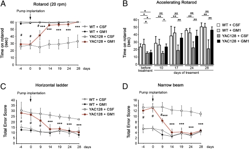
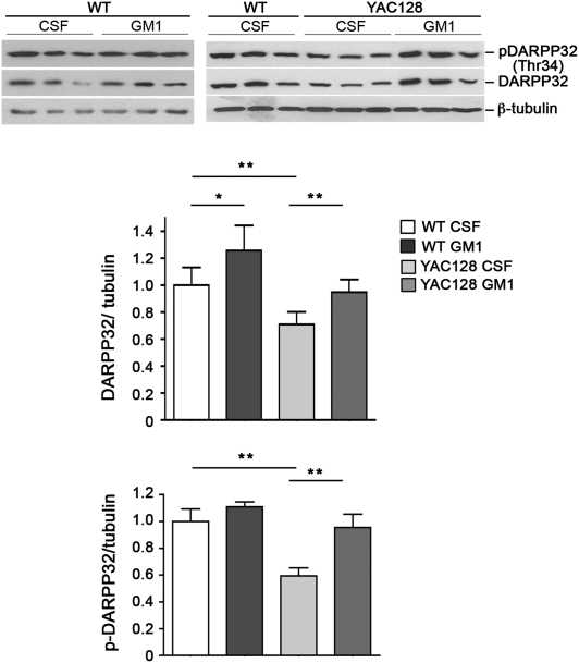
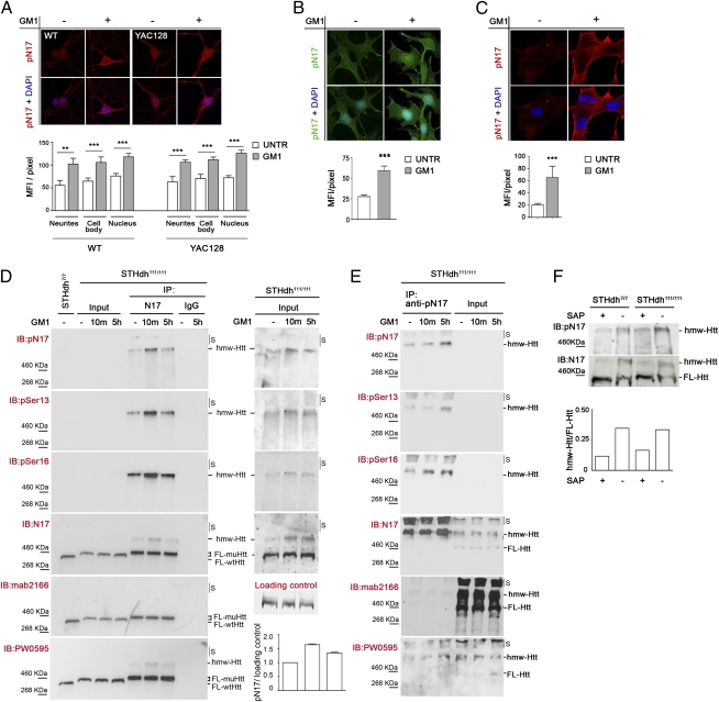
Huntington disease (HD) is a progressive neurodegenerative monogenic disorder caused by expansion of a polyglutamine stretch in the huntingtin (Htt) protein. Mutant huntingtin triggers neural dysfunction and death, mainly in the corpus striatum and cerebral cortex, resulting in pathognomonic motor symptoms, as well as cognitive and psychiatric decline. Currently, there is no effective treatment for HD. We report that intraventricular infusion of ganglioside GM1 induces phosphorylation of mutant huntingtin at specific serine amino acid residues that attenuate huntingtin toxicity, and restores normal motor function in already symptomatic HD mice. Thus, our studies have identified a potential therapy for HD that targets a posttranslational modification of mutant huntingtin with critical effects on disease pathogenesis.


Conflict of interest statement

The authors declare no conflict of interest.

Figures

Fig. 1.
Fig. 1.
GM1 restores normal motor behavior in YAC128 mice. Behavioral tests were conducted at the indicated time, on 5-mo-old YAC128 and WT mice, before and during GM1 chronic brain infusion. Each data point represents the average performance ±SD of six mice. (A) Rotarod test at fixed speed (20 rpm for 60 s). YAC128 mice treated with GM1 showed progressive improvement and, by the end of the treatment, were able to finish the test like most WT mice. The horizontal gray line in the graph marks the test endpoint. (B) Accelerating rotarod (4–40 rpm in 1 min). In this challenging test, YAC128 mice treated with GM1 performed as well as WT mice (differences between WT and GM1-treated YAC128 mice were not statistically significant). (C) Horizontal ladder test. The ability of mice to cross a horizontal ladder with irregular rung pattern was analyzed. A score was assigned to each type of footfall and other mistakes made by the mice according to ref. . (D) Narrow beam test. Motor performance was scored as the mice walked along a narrow beam (100 cm long, 0.75 cm wide). #P < 0.01 (YAC128 vs. WT); *P < 0.05; **P < 0.01; ***P < 0.001 (GM1-treated vs. CSF-treated YAC128).
Fig. 2.
Fig. 2.
GM1 restores striatal expression of DARPP-32 in YAC128 mice. Representative immunoblot and Li-Cor Odissey densitometric analysis of DARPP-32 expression and activation (p-DARPP-32) in WT and YAC128 mice. GM1 infusion for 28 d restores normal levels of DARPP-32 and active p-DARPP-32 in YAC128 mice. Each lane of the immunoblot represents one individual mouse. Graphs show the densitometric analysis performed on three (WT GM1) or six (all other experimental groups) mice per group. Bars represent the mean values ±SD *P < 0.05; **P < 0.01.
Fig. 3.
Fig. 3.
GM1 administration elicits huntingtin phosphorylation at serine 13 and serine 16. (A) Representative confocal microscopy images and relative quantitative analysis of primary striatal neurons isolated from wild-type (WT) and YAC128 mice and incubated for 5 h with 50 μM GM1 (+) or vehicle (−). Neurons were immunostained with anti–phospho-N17 (pN17) antibody, which recognizes the amino-terminal N17 peptide of huntingtin phosphorylated at amino acid residues serine 13 and serine 16, and with DAPI to visualize nuclei. (B) Representative epifluorescence microscope images and quantitative analysis of immortalized knock-in striatal progenitor cells expressing mutant huntingtin (STHdh111/111) and treated as in A. (C) Confocal microscopy images and quantitative analysis of primary fibroblasts from HD patients treated as in A. Graphs in AC show pN17 immunostaining mean fluorescence intensity (MFI) per pixel ±SD, calculated over a minimum of 100 cells per experimental group. **P < 0.01; ***P < 0.001. (D and E) Analysis of mutant huntingtin phosphorylation state by immunoprecipitation and immunoblotting. Striatal knock-in cells (STHdh111/111) were incubated with 50 μM GM1 for the indicated time (10m, 10 min). Mutant huntingtin was immunoprecipitated from equal amounts of total cell lysates using a rabbit polyclonal anti-huntingtin antibody (N17) (in D), phospho-specific pN17 antibodies (in E), or nonspecific rabbit IgG antibodies as negative control (D). Total lysate from cells expressing wild-type huntingtin (STHdh7/7) was loaded in the same gel as reference. All immunoprecipitated material (IP) was immunoblotted with the indicated phospho-specific and anti-huntingtin antibodies. Phosphohuntingtin could not be detected in the total lysates (input lanes, 30 μg of proteins loaded) due to the proximity of the highly immune-reactive immunoprecipitated material in adjacent lanes. The results of reprobing the input lanes only, after cutting the membrane, are shown in D, Right. An increase in the phosphorylation of huntingtin at serine 13 and serine 16 after treatment with GM1 is evident in both immunoprecipitated material and input total cell lysates. The graph in D shows the densitometric analysis of huntingtin phosphorylation in the input lysates of two independent experiments. A Ponceau red-stained band in the membrane was used as loading control. S, stacking portion of the gel. (F) Wild-type and mutant huntingtin in total cell lysate from striatal knock-in cells (STHdh7/7 and STHdh111/111) was dephosphorylated using shrimp alkaline phosphatase (SAP). Dephosphorylation resulted in a dramatic change in huntingtin electrophoretic mobility, with increased amount of protein migrating at the lower apparent molecular weight (FL-Htt) and a decreased amount of high molecular-weight species (hmw-Htt). The graph shows the ratio between hmw-Htt and FL-Htt before and after dephosphorylation with SAP.
Fig. 4.
Fig. 4.
GM1 infusion induces huntingtin phosphorylation in vivo. Total protein lysates were prepared from the cortex and the striatum of YAC128 mice and WT littermates chronically infused with artificial cerebrospinal fluid (CSF) or GM1 in CSF for 28 d (six mice per group). (A) Representative immunoblots showing increased phosphorylation of huntingtin at serine 13 and serine 16 in GM1-infused brains, as detected using pN17 antibody. Each lane corresponds to the lysate from one mouse. (B) Li-Cor Odyssey infrared densitometric analysis of phosphohuntingtin normalized over talin (loading control). Bars represent the mean ± SD of six mice per experimental group. *P < 0.05; ***P < 0.001.

References

    1. Zuccato C, Valenza M, Cattaneo E. Molecular mechanisms and potential therapeutical targets in Huntington's disease. Physiol Rev. 2010;90:905–981. - PubMed
    1. Steffan JS, et al. SUMO modification of Huntingtin and Huntington's disease pathology. Science. 2004;304:100–104. - PubMed
    1. Graham RK, et al. Cleavage at the caspase-6 site is required for neuronal dysfunction and degeneration due to mutant huntingtin. Cell. 2006;125:1179–1191. - PubMed
    1. Jeong H, et al. Acetylation targets mutant huntingtin to autophagosomes for degradation. Cell. 2009;137:60–72. - PMC - PubMed
    1. Schilling B, et al. Huntingtin phosphorylation sites mapped by mass spectrometry. Modulation of cleavage and toxicity. J Biol Chem. 2006;281:23686–23697. - PubMed

Publication types

MeSH terms

LinkOut - more resources