. 2012 Mar 6;109(10):3921-6.
doi: 10.1073/pnas.1117135109. Epub 2012 Feb 13.

High-grade serous ovarian cancer arises from fallopian tube in a mouse model

Affiliations

High-grade serous ovarian cancer arises from fallopian tube in a mouse model

Jaeyeon Kim et al. Proc Natl Acad Sci U S A. .

Abstract

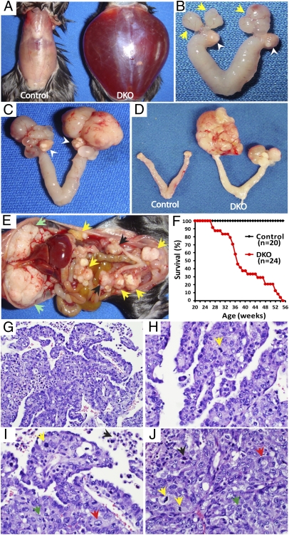
Although ovarian cancer is the most lethal gynecologic malignancy in women, little is known about how the cancer initiates and metastasizes. In the last decade, new evidence has challenged the dogma that the ovary is the main source of this cancer. The fallopian tube has been proposed instead as the primary origin of high-grade serous ovarian cancer, the subtype causing 70% of ovarian cancer deaths. By conditionally deleting Dicer, an essential gene for microRNA synthesis, and Pten, a key negative regulator of the PI3K pathway, we show that high-grade serous carcinomas arise from the fallopian tube in mice. In these Dicer-Pten double-knockout mice, primary fallopian tube tumors spread to engulf the ovary and then aggressively metastasize throughout the abdominal cavity, causing ascites and killing 100% of the mice by 13 mo. Besides the clinical resemblance to human serous cancers, these fallopian tube cancers highly express genes that are known to be up-regulated in human serous ovarian cancers, also demonstrating molecular similarities. Although ovariectomized mice continue to develop high-grade serous cancers, removal of the fallopian tube at an early age prevents cancer formation--confirming the fallopian tube origin of the cancer. Intriguingly, the primary carcinomas are first observed in the stroma of the fallopian tube, suggesting that these epithelial cancers have a mesenchymal origin. Thus, this mouse model demonstrates a paradigm for the origin and initiation of high-grade serous ovarian carcinomas, the most common and deadliest ovarian cancer.


Conflict of interest statement

The authors declare no conflict of interest.

Figures

Fig. 1.
Fig. 1.
Dicer-Pten DKO mice develop high-grade metastatic serous carcinoma. (A) Severe ascites in an 8.4-mo-old DKO mouse. (B) Early tumors form in the fallopian tube (yellow arrows) of a 5-mo-old DKO mouse with normal ovaries (white arrowheads). (C) Progression of the fallopian tube tumors in a DKO mouse at 8 mo. Ovaries are still intact (white arrowheads). (D) Bilateral fallopian tube/ovarian tumors are observed in a DKO mouse at 6 mo. (E) The DKO mouse described in A showing extensive peritoneal metastasis with clusters of tumor nodules (yellow arrows) and a massive accumulation on the diaphragm (green arrows), besides fallopian tube/ovarian tumors (black arrows). (F) Survival curve of DKO and control mice. (GI) DKO fallopian tube/ovarian tumors showing papillary structure and irregular glands with slit-like spaces, characteristic of high-grade serous carcinoma (H&E) and representative of fallopian tube/ovarian tumors from 16 DKO mice. (HJ) The nuclear features of high-grade serous carcinomas including nuclear pleomorphism (red arrow), prominent nucleoli with irregular chromatin patterns (green arrow), apoptosis (black arrow), and brisk mitotic activity (yellow arrow) (H&E). (J) Fallopian tube/ovarian tumor with a solid growth pattern (H&E). Magnifications: G, 20×; H–J, 40×.)
Fig. 2.
Fig. 2.
Molecular alterations in Dicer-Pten DKO mice. (A) Western blot analysis of DKO fallopian tube tumors showing activation of PI3K signaling compared with control fallopian tubes as indicated by the enhanced expression of phosphorylated (P)-AKT, P-PRAS40 (AKT1S1), P-4EBP1, survivin, and stathmin. On the right side, mRNA enrichment (fold change) in the mouse fallopian tube cancers versus control fallopian tubes is presented. (B) DNA copy number changes in the PTEN and DICER1 alleles in 481 high-grade serous ovarian tumors from The Cancer Genome Atlas (TCGA): yellow, gain; blue, loss. Values are from Affymetrix SNP 1M array dataset.
Fig. 3.
Fig. 3.
Fallopian tube is the origin of high-grade serous carcinomas in Dicer-Pten DKO mice. (A) Tumor (black arrowhead) in an 8-mo-old DKO mouse after unilateral removal of the ovary. (B) No tumors in the ovary (white arrowhead) in an 8-mo-old DKO mouse with the fallopian tube removed unilaterally. (C) After removal of both ovaries, massive tumors still form from the fallopian tubes in a 10.5-mo-old DKO mouse. (D) No tumors with both fallopian tubes removed in an 11.5-mo DKO mouse. (E) Histology of an early fallopian tube lesion displaying high-grade serous carcinomas (representative of early tumors from seven DKO mice), in which tumor cells extensively infiltrate and expand the stroma of the fallopian tube (H&E). (F and G) Proliferating tumor cells show strong and abundant immunohistochemical staining of cytokeratin 14 (KRT14) and cytokeratin 8 (KRT8). (H) Tumor cells are primarily located within the stroma with preservation (arrow) and focal attenuation (arrowhead) of overlying benign-looking tubal epithelium (H&E, 20× magnification of the dotted region from E). (I) KRT14-positive tumor cells focally invade and erode the fallopian tube epithelium (20× magnification of the dotted region from F). (J) Proliferating tumor cells show abundant Ki67 expression with no significant expression in fallopian tube epithelium (arrowheads). (K) Abundant CDH1 expression in tumor cells of fallopian tube stroma (arrow) and in the epithelial layer (arrowheads). (L and M) An early fallopian tube lesion. A small nest (long arrow) and a few single tumor cells (short arrow) show strong KRT14 (L) and KRT17 (M) expression, compared with fallopian tube epithelium (arrowhead) and uninvolved stroma that are KRT14-negative (L). (N) Specific CA125 staining in these early tumor clusters (long arrow) in the fallopian tube stroma. (O and P) Histology of deeper sections obtained from the early tubal lesion corresponding to LN. High-grade carcinoma cells form nests and ill-defined glands (dotted circles), which are primarily expanding the stroma. There is focal involvement of the serosal surface (O, arrow) with essentially uninvolved tubal epithelium in early lesions (P, arrowhead). (Scale bars: 0.5 cm.) (Magnification: EG, 4×; H and I, 20×; JL, 10×; MP, 20×.)

Comment in

  • Nonovarian origins of ovarian cancer.
    Hillier SG. Hillier SG. Proc Natl Acad Sci U S A. 2012 Mar 6;109(10):3608-9. doi: 10.1073/pnas.1201029109. Epub 2012 Feb 22. Proc Natl Acad Sci U S A. 2012. PMID: 22357760 Free PMC article. No abstract available.

References

    1. Cho KR, Shih IeM. Ovarian cancer. Annu Rev Pathol. 2009;4:287–313. - PMC - PubMed
    1. Koonings PP, Campbell K, Mishell DR, Jr, Grimes DA. Relative frequency of primary ovarian neoplasms: A 10-year review. Obstet Gynecol. 1989;74:921–926. - PubMed
    1. Seidman JD, et al. The histologic type and stage distribution of ovarian carcinomas of surface epithelial origin. Int J Gynecol Pathol. 2004;23:41–44. - PubMed
    1. Jemal A, et al. Cancer statistics, 2009. CA Cancer J Clin. 2009;59:225–249. - PubMed
    1. Bukowski RM, Ozols RF, Markman M. The management of recurrent ovarian cancer. Semin Oncol. 2007;34(Suppl 2):S1–S15. - PubMed

Publication types