. 2012 Feb 28;109(9):3451-6.
doi: 10.1073/pnas.1201042109. Epub 2012 Feb 13.

Contribution of IL-33-activated type II innate lymphoid cells to pulmonary eosinophilia in intestinal nematode-infected mice

Affiliations

Contribution of IL-33-activated type II innate lymphoid cells to pulmonary eosinophilia in intestinal nematode-infected mice

Koubun Yasuda et al. Proc Natl Acad Sci U S A. .

Abstract

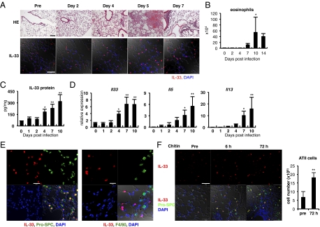
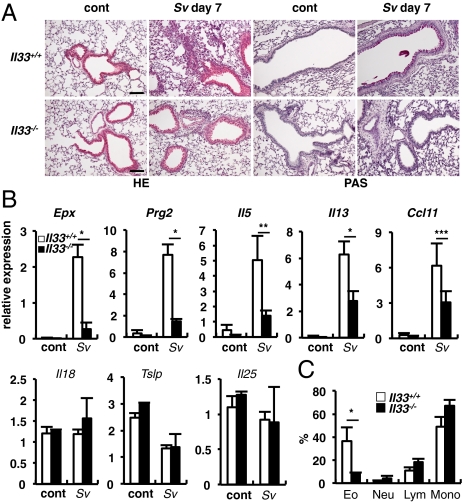
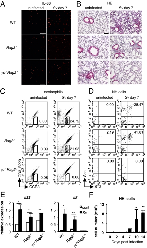
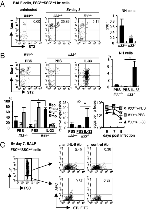
When animals are infected with helminthic parasites, resistant hosts show type II helper T immune responses to expel worms. Recently, natural helper (NH) cells or nuocytes, newly identified type II innate lymphoid cells, are shown to express ST2 (IL-33 receptor) and produce IL-5 and IL-13 when stimulated with IL-33. Here we show the relevant roles of endogenous IL-33 for Strongyloides venezuelensis infection-induced lung eosinophilic inflammation by using Il33(-/-) mice. Alveolar epithelial type II cells (ATII) express IL-33 in their nucleus. Infection with S. venezuelensis or intranasal administration of chitin increases in the number of ATII cells and the level of IL-33. S. venezuelensis infection induces pulmonary accumulation of NH cells, which, after being stimulated with IL-33, proliferate and produce IL-5 and IL-13. Furthermore, S. venezuelensis infected Rag2(-/-) mice increase the number of ATII cells, NH cells, and eosinophils and the expression of IL-33 in their lungs. Finally, IL-33-stimulated NH cells induce lung eosinophilic inflammation and might aid to expel infected worms in the lungs.


Conflict of interest statement

The authors declare no conflict of interest.

Figures

Fig. 1.
Fig. 1.
S. venezuelensis infection increases IL-33 expression in the lungs. (A–E) WT mice were infected with S. venezuelensis at day 0. (A) Histological analysis of lungs was performed at indicated days. (Upper) HE, stained with H&E. (Scale bar, 100 μm.) (Lower) Confocal microscopic analysis of the IL-33 expression. Red, IL-33; blue, DAPI. (Scale bar, 50 μm.) (B) The number of eosinophils in BALFs at indicated days (n = 3 ∼4). (C and D) IL-33 concentration in the lung lysates was examined by ELISA. The amounts of IL-33 were normalized by the total protein concentration (C). Quantitative RT-PCR (qPCR) analysis of the expression levels of mRNA for Il33, Il5, or Il13 in the lungs (D). Data are representative of two independent experiments and expressed as the means ± SD (n = 5) *P < 0.01, **P < 0.001 (B–D; one-way ANOVA with Dunnett's post test). (E) IL-33 (red) in the lungs at day 7 postinfection was costained with pro-SPC (green, Left) or F4/80 (green, Right). Blue, DAPI. (Scale bars, 20 μm.) (F) Chitin was intranasally administered into B6 mice. (Left) Confocal microscopic analysis of the lungs at indicated time points. Red, IL-33; blue, DAPI; green, pro-SPC. (Scale bar, 50 μm.) (Right) Lung cells were prepared from nontreated mice or mice treated with chitin 72 h before (n = 3). The numbers of ATII cells were calculated as described in Fig. S3.
Fig. 2.
Fig. 2.
Il33−/− mice showed reduced accumulations of eosinophils in the lungs after S. venezuelensis infection. (A) Histological analysis of lungs of Il33+/+ and Il33−/− was performed before (cont) and after S. venezuelensis infection (Sv). PAS, periodic acid-Schiff stain. (Scale bars, 100 μm.) (B) Total RNA was prepared from lungs and the levels of mRNA expressions for Epx, Prg2 and indicated cytokines were determined by qPCR. Data are representative of two independent experiments and expressed as the means ± SD (n = 3 ∼5) *P < 0.01, **P < 0.001, ***P < 0.05 (Student's t test). (C) Proportions of eosinophils (Eo), neutrophils (Neu), lymphocytes (Lym), or monocytes (Mono) in the BALF were calculated from flow cytometric analysis of CD45+ BALF cells from S. venezuelensis infected mice (Sv). Data are representative of two independent experiments and expressed as the means ± SD (n = 3 ∼5) *P < 0.05, (Student's t test).
Fig. 3.
Fig. 3.
WT, Rag2−/−, and γc−/−Rag2−/− (n = 3~5) mice were uninfected (cont) or infected with S. venezuelensis (Sv). (A) Confocal microscopic analysis of the IL-33 (red) expression in the lungs. (Scale bars, 50 μm.) (B) Histological analysis (H&E) of lungs. (Scale bar, 100 μm.) (C and D) Flow cytometric analysis of the BALF cells. Numbers indicate proportion of eosinophils (C) or NH cells (D). Cells were gated on the CD45+ fraction (C) or FSClowSSClowLin fraction (D). (E) qPCR analysis of the expression levels of mRNA for Il33 and Il5 in the lungs. Data are expressed as the means ± SD *P < 0.01, **P < 0.05 (Student's t test). (F) The numbers of NH cells in the BALFs from mice in Fig. 1B are shown. Data are expressed as the means ± SD *P < 0.01, **P < 0.001 (one-way ANOVA with Dunnett's post test). Data are representative of two independent experiments.
Fig. 4.
Fig. 4.
IL-33-dependent induction of NH cells in S. venezuelensis infected mice. (A) Flow cytometric analysis of NH cells in BALF cells from uninfected or S. venezuelensis infected (Sv) Il33+/+ or Il33−/− mice. Cells were gated on the FSClowSSClowLin fraction. (Right) The numbers of NH cells in total BALF cells from Il33+/+ (n = 11) or Il33−/− (n = 5) mice. Data are expressed as the means ± SD *P < 0.0005 (Student's t test). (B) Il33+/+ (n = 7) and Il33−/− mice (PBS; n = 4, IL-33; n = 6) were infected with S. venezuelensis at day 0 and treated daily with 30 μL of PBS or 4 μg rhIL-33 intranasally from day −1 to 3. (Upper) Flow cytometry of NH cells in FSClowSSClowLin fraction of BALF cells and the proportion of NH cells in total BALF cells at 8 d postinfection. (Lower Left) The proportion of eosinophils (eo), neutrophils (neu), lymphocytes (lym), or monocytes (mo) in BALF cells. (Lower Center) The expression of Il5 mRNA in the lungs. (Lower Right) The numbers of eggs per gram feces from each group at day 6, 7, or 8 postinfection. Data are expressed as the means ± SD; *P < 0.001, **P < 0.05 versus corresponding values for Il33−/− mice with PBS treatment (Student's t test). (C) Intracellular staining of IL-5 in LinST2+ cells in BALF cells. Cells were stained as described in SI Materials and Methods. Numbers indicate the proportion of ST2+IL-5+ cells. Data are representative of five mice and of two independent experiments.

References

    1. Finkelman FD, et al. Interleukin-4- and interleukin-13–mediated host protection against intestinal nematode parasites. Immunol Rev. 2004;201:139–155. - PubMed
    1. Maizels RM, Pearce EJ, Artis D, Yazdanbakhsh M, Wynn TA. Regulation of pathogenesis and immunity in helminth infections. J Exp Med. 2009;206:2059–2066. - PMC - PubMed
    1. Nakanishi K. Basophils are potent antigen-presenting cells that selectively induce Th2 cells. Eur J Immunol. 2010;40:1836–1842. - PubMed
    1. Paul WE, Zhu J. How are T(H)2-type immune responses initiated and amplified? Nat Rev Immunol. 2010;10:225–235. - PMC - PubMed
    1. Matsuba-Kitamura S, et al. Contribution of IL-33 to induction and augmentation of experimental allergic conjunctivitis. Int Immunol. 2010;22:479–489. - PubMed

Publication types

MeSH terms

LinkOut - more resources