

Image quality of digital direct flat-panel mammography versus an indirect small-field CCD technique using a high-contrast phantom
- PMID: 22332015
- PMCID: PMC3276250
- DOI: 10.4061/2011/701054
Image quality of digital direct flat-panel mammography versus an indirect small-field CCD technique using a high-contrast phantom
Abstract
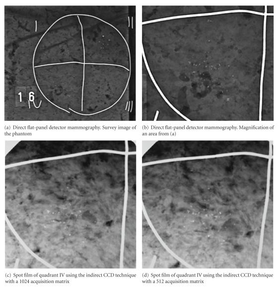
Objective: To compare the detection of microcalcifications on mammograms of an anthropomorphic breast phantom acquired by a direct digital flat-panel detector mammography system (FPM) versus a stereotactic breast biopsy system utilizing CCD (charge-coupled device) technology with either a 1024 or 512 acquisition matrix (1024 CCD and 512 CCD).
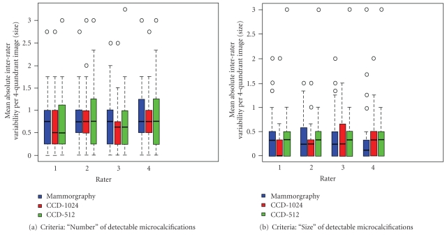
Materials and methods: Randomly distributed silica beads (diameter 100-1400 μm) and anthropomorphic scatter bodies were applied to 48 transparent films. The test specimens were radiographed on a direct digital FPM and by the indirect 1024 CCD and 512 CCD techniques. Four radiologists rated the monitor-displayed images independently of each other in random order.
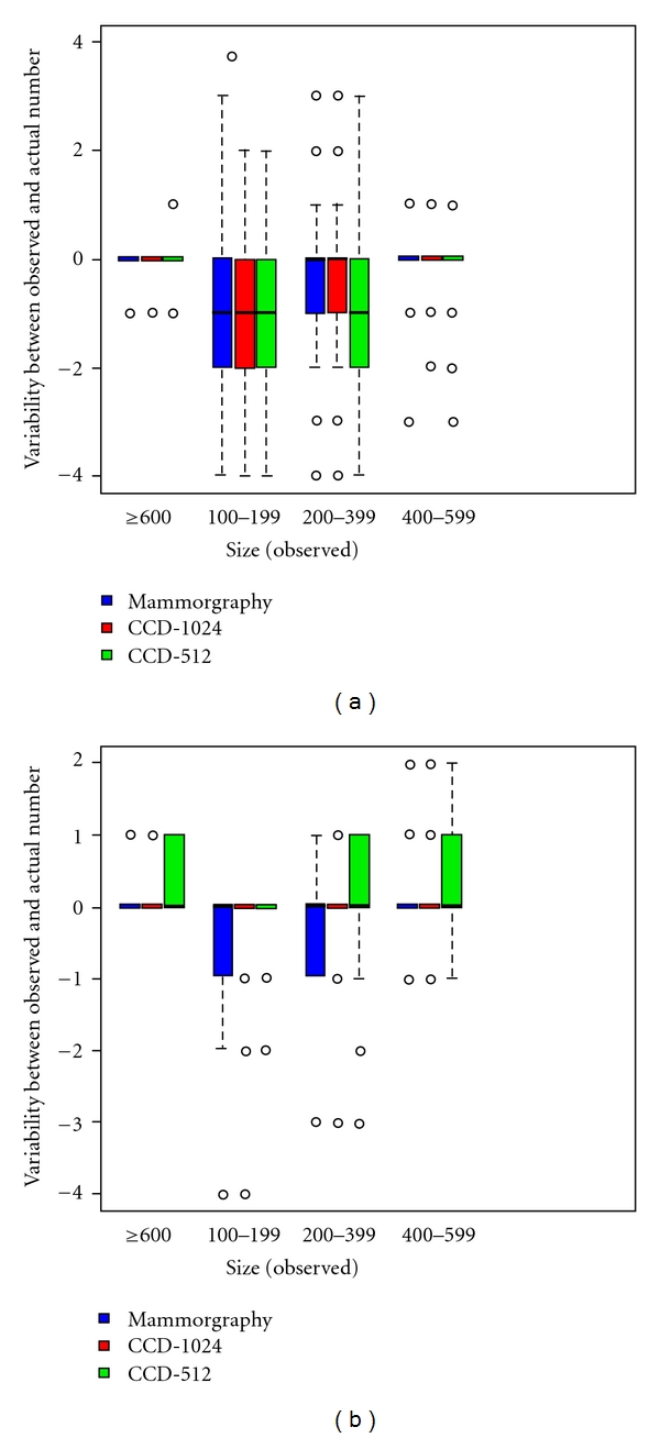
Results: The rate of correct positive readings for the "number of detectable microcalcifications" for silica beads of 100-199 μm in diameter was 54.2%, 50.0% and 45.8% by FPM, 1024 CCD and 512 CCD, respectively. The inter-rater variability was most pronounced for silica beads of 100-199 μm in diameter. The greatest agreement with the gold standard was observed for beads >400 μm in diameter across all methods.
Conclusion: Stereotactic spot images taken by 1024 matrix CCD technique are diagnostically equivalent to direct digital flat-panel mammograms for visualizing simulated microcalcifications >400 μm in diameter.
Figures
Similar articles
-
Image quality of digital direct flat-panel mammography versus an analog screen-film technique using a phantom model.AJR Am J Roentgenol. 2007 Feb;188(2):399-407. doi: 10.2214/ajr.05.2006. AJR Am J Roentgenol. 2007. PMID: 17242248
-
Image quality of digital direct flat-panel mammography versus an analog screen-film technique using a low-contrast phantom.AJR Am J Roentgenol. 2008 Sep;191(3):W80-8. doi: 10.2214/AJR.07.2870. AJR Am J Roentgenol. 2008. PMID: 18716083
-
Comparison of the Detection Rate of Simulated Microcalcifications in Full-Field Digital Mammography, Digital Breast Tomosynthesis, and Synthetically Reconstructed 2-Dimensional Images Performed With 2 Different Digital X-ray Mammography Systems.Invest Radiol. 2017 Apr;52(4):206-215. doi: 10.1097/RLI.0000000000000334. Invest Radiol. 2017. PMID: 27861206
-
Quantification of Al-equivalent thickness of just visible microcalcifications in full field digital mammograms.Med Phys. 2004 Jul;31(7):2165-76. doi: 10.1118/1.1758352. Med Phys. 2004. PMID: 15305471
-
Visibility of simulated microcalcifications--a hardcopy-based comparison of three mammographic systems.Med Phys. 2005 Jan;32(1):182-94. doi: 10.1118/1.1833011. Med Phys. 2005. PMID: 15719969
Cited by
-
Cancelled stereotactic biopsy of calcifications not seen using the stereotactic technique: do we still need to biopsy?Eur Radiol. 2014 Apr;24(4):907-12. doi: 10.1007/s00330-013-3055-z. Epub 2013 Nov 12. Eur Radiol. 2014. PMID: 24217642
References
-
- Kreienberg R, Kopp I, Lorenz W, et al. Interdisziplinäre Leitlinie der Deutschen Krebsgesellschaft und der beteiligten medizinisch-wissenschaft-lichen Fachgesellschaften. Diagnostik, Therapie und Nachsorge des Mammakarzinoms der Frau. Eine nationale S-3-Leitlinie. http://www.uni-duesseldorf.de/WWW/AWMF/ll/032-045.pdf.
-
- Perry N, Broeders M, de Wolf F, Törnberg S, Holland R, von Karsa L, editors. European Guidelines for Quality Assurance in Breast Cancer Screening and Diagnosis. 4th edition. Köln, Germany: European Commission, Bundesanzeiger Verlag GmbH; 2006. http://www.euref.org. - PubMed
-
- Ciatto S, Houssami N, Ambrogetti D, et al. Accuracy and underestimation of malignancy of breast core needle biopsy: the Florence experience of over 4000 consecutive biopsies. Breast Cancer Research and Treatment. 2007;101(3):291–297. - PubMed
-
- Diekmann F, Diekmann S, Bick U, et al. Comparing the visualization of microcalcifications with direct magnification in digital full-field mammography vs. film-screen mammography. Fortschr Röntgenstr. 2002;174(3):297–300. - PubMed
-
- Diekmann S, Bick U, Von Heyden H, Diekmann F. Visualization of microcalcifications on mammographies obtained by digital fullfield mammography in comparison to conventional film-screen mammography. Fortschr Röntgenstr. 2003;175(6):775–779. - PubMed
LinkOut - more resources
Full Text Sources
