. 2010 Mar;23(1):43-68.
doi: 10.1089/ped.2010.0010.

Imaging of Childhood Interstitial Lung Disease

Affiliations

Imaging of Childhood Interstitial Lung Disease

R Paul Guillerman. Pediatr Allergy Immunol Pulmonol. 2010 Mar.

Abstract

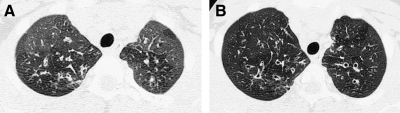
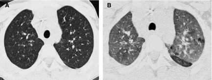
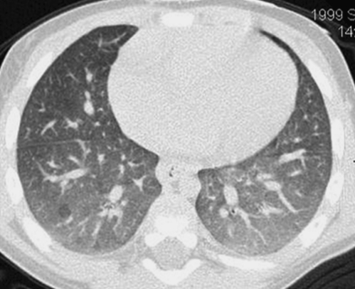
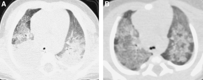
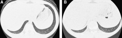
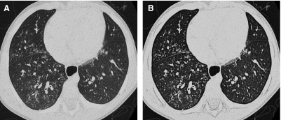
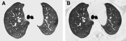
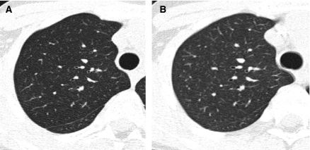
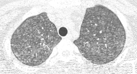
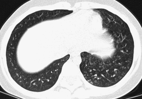
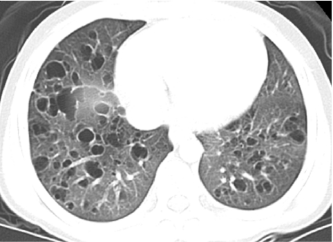
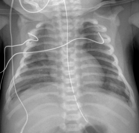
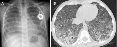
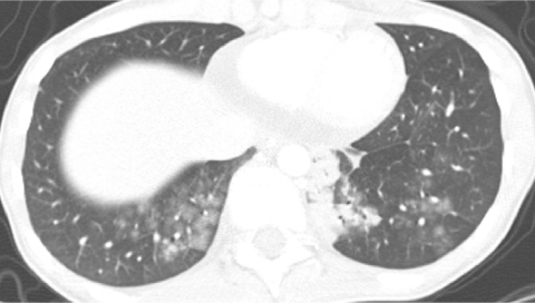
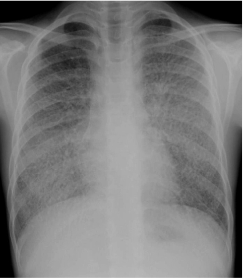
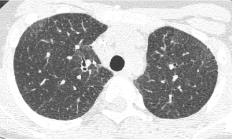
The aphorism that children are not little adults certainly applies for the imaging of interstitial lung disease. Acquiring motion-free images of fine pulmonary structures at desired lung volumes is much more difficult in children than in adults. Several forms of interstitial lung disease are unique to children, and some forms of interstitial lung disease encountered in adults rarely, if ever, occur in children. Meticulous attention to imaging technique and specialized knowledge are required to properly perform and interpret chest imaging studies obtained for the evaluation of childhood interstitial lung disease (chILD). This review will address technique recommendations for imaging chILD, the salient imaging findings in various forms of chILD, and the efficacy of imaging in the diagnosis and management of chILD.


Figures

FIG. 1.
FIG. 1.
Fine anatomic details of the lungs are much better depicted on a thin (1.25 mm) axial image slice (A) from an HRCT exam than on a thicker (5 mm) axial image slice (B) from a routine chest CT exam obtained at the same level.
FIG. 2.
FIG. 2.
Volumetric multislice CT technique covers the entire lungs (A). Conventional noncontiguous HRCT technique samples only portions of the lungs at increments (B), leaving gaps that are not imaged, but exposing the patient to a lower radiation dose.
FIG. 3.
FIG. 3.
A thin-section CT image reconstructed from volumetric data acquired by modern multislice helical CT technique (A) is very similar in quality to an image acquired by conventional noncontiguous HRCT technique (B).
FIG. 4.
FIG. 4.
Blurring of pulmonary vessels from motion induced by transmitted cardiac pulsations can obscure or simulate pathology such as bronchiectasis on CT, particularly in the lower left lung adjacent to the heart.
FIG. 5.
FIG. 5.
The fuller the lung inflation the greater the distention of the airways; consequently, bronchiectasis that is occult on HRCT performed at low lung volumes (A) may become overt on HRCT performed at deep inspiration (B).
FIG. 6.
FIG. 6.
Compared with its appearance on inspiratory HRCT (A), the normal lung manifests increased attenuation on expiratory HRCT (B) due to the lower proportion of air relative to solid pulmonary parenchymal tissue at smaller lung volumes. An inspiratory state is suggested by a convex contour of the posterior tracheal membrane, while a horizontal or concave contour of the posterior tracheal membrane suggests an expiratory state.
FIG. 7.
FIG. 7.
A ground-glass appearance with hazy increased attenuation of the lungs and preserved visibility of the bronchovascular structures can occur physiologically on a chest CT image obtained at tidal volumes during quiet respiration and simulate diffuse lung disease. This physiologic increase in lung attenuation is more pronounced dependently where the pulmonary vascular blood volume is greatest and the airspaces are least distended (image courtesy of Fred Long, M.D.).
FIG. 8.
FIG. 8.
A CT image shows confluent opacification of the dependent posterior aspects of the lungs of an infant scanned while supine (A). A CT image acquired after prone repositioning of the patient (B) re-expands the posterior atelectasis and reveals underlying patchy pulmonary consolidation and ground-glass opacification related to surfactant dysfunction from an SP-C gene mutation.
FIG. 9.
FIG. 9.
A supine chest CT image from a teenager with systemic sclerosis image shows peripheral opacities at the dependent poster-obasilar lower lobes (A). A CT image acquired after prone repositioning of the patient reveals persistence of these opacities which represent pulmonary involvement by systemic sclerosis rather than just atelectasis (B).
FIG. 10.
FIG. 10.
An HRCT image generated with a “bone” reconstruction algorithm (A) is preferable to an HRCT image generated with a “lung” reconstruction algorithm (B) due to excessive edge enhancement artifact with the latter, exemplified by the dark etching at the lung periphery.
FIG. 11.
FIG. 11.
A hypoattenuating focus of air trapping at the peripheral lateral left upper lobe is more conspicuous on an HRCT image viewed with a narrow (900 HU) window width (A) than with a conventional (1,500 HU) window width (B), but the narrow window width produces artifactual bronchial wall thickening.
FIG. 12.
FIG. 12.
There is no clinically relevant difference in the diagnostic quality of this HRCT image (A) compared with an HRCT image obtained with a 3 times greater radiation dose (B). The inherent high contrast between air and lung soft tissues permits HRCT images of diagnostic quality to be acquired with relatively low radiation doses, particularly if the lungs are well inflated.
FIG. 13.
FIG. 13.
Image from an infant HRCT obtained with an estimated radiation effective dose of 0.1 milliSievert, which is equivalent to the dose from a few chest radiographs (CXRs). Although the image quality is degraded by the high noise level, the pattern of ground-glass opacities is sufficiently depicted to corroborate the clinical diagnosis of neuroendocrine cell hyperplasia of infancy.
FIG. 14.
FIG. 14.
Inspiratory (A) and expiratory (B) HRCT images of a patient with bronchiolitis obliterans show mosaic attenuation of the lungs with patchy ground-glass opacification that is accentuated on expiration. Areas of pulmonary air trapping related to obliterated airways exhibit decreased conspicuity of the pulmonary vessels, little decrease in volume on expiration, and little increase in attenuation on expiration. Areas of spared normal lung exhibit marked increased attenuation on expiration related to the decreased volume of air in the airspaces and the increased capillary blood flow diverted from the areas with air trapping.
FIG. 15.
FIG. 15.
Chest CT image from a case of aspiration pneumonia showing consolidation with air bronchograms involving the lower lobes of the lungs.
FIG. 16.
FIG. 16.
HRCT in a patient with lymphangiomatosis shows linear and polygonal opacities related to interlobular septal thickening, most conspicuously at the peripheral paramediastinal and posterior lung regions.
FIG. 17.
FIG. 17.
A fine reticular pattern of septal thickening is demonstrated on this HRCT image from a child with nonspecific interstitial pneumonia.
FIG. 18.
FIG. 18.
A chest CT image from a patient with juvenile dermatomyositis shows coarse peripheral opacities with subpleural bands, architectural distortion, and traction bronchiectasis related to pulmonary fibrosis.
FIG. 19.
FIG. 19.
HRCT image from a patient with acute myelogenous leukemia and idiopathic pneumonia syndrome shows a “crazy-paving” pattern of superimposed septal thickening and ground-glass opacification, most conspicuously in the right lung.
FIG. 20.
FIG. 20.
The presence of perilymphatic nodules gives a beaded appearance to some of the bronchovascular bundles on this chest CT image from a patient with pulmonary sarcoidosis.
FIG. 21.
FIG. 21.
Hematogenous dissemination of the disease process in miliary tuberculosis is represented by the presence of randomly distributed pulmonary nodules on this chest CT image.
FIG. 22.
FIG. 22.
Numerous centrilobular ground-glass nodular opacities are depicted on this chest CT image from an adolescent with hypersensitivity pneumonitis.
FIG. 23.
FIG. 23.
Branching centrilobular opacities in a tree-in-bud pattern are shown in the left lower lobe on this chest CT image from an adolescent with an atypical mycobacterial infection and lymphocytic bronchiolitis.
FIG. 24.
FIG. 24.
A chest CT image from a child with pulmonary Langerhans cell histiocytosis demonstrates numerous air-filled thin-walled cysts in the lungs.
FIG. 25.
FIG. 25.
CXR of a full-term neonate with alveolar capillary dysplasia with misalignment of the pulmonary veins shows hazy pulmonary opacities resembling the findings of surfactant deficiency. Small pneumothoraces and pneumomediastinum related to air leak from barotrauma are also present.
FIG. 26.
FIG. 26.
A chest CT image from a very premature neonate with bronchopulmonary dysplasia exhibits hyperlucent areas that correspond to alveolar enlargement and simplification and reduced distal vascularization, and linear opacities that correspond to interstitial fibroproliferation.
FIG. 27.
FIG. 27.
A chest CT image from a 4-month-old term infant with respiratory insufficiency showing distortion of the lung architecture with perilobular opacities and pulmonary lobules of variable attenuation. Histopathologic inspection revealed a lung growth disorder characterized by lobular simplification and alveolar enlargement.
FIG. 28.
FIG. 28.
Multiple subpleural cysts are demonstrated on this chest CT image from a 5-year-old with a lung growth disorder associated with trisomy 21.
FIG. 29.
FIG. 29.
An HRCT image from an infant with a filamin A gene mutation shows severe multilobar pulmonary hyperinflation and hyperlucency with peripheral pulmonary vascular attenuation resembling emphysema. A lung growth disorder characterized by simplified enlarged alveolar airspaces and pulmonary hypertensive changes was noted on a lung biopsy specimen.
FIG. 30.
FIG. 30.
Chest CT images of a 3-month-old infant with persistent tachypnea show mosaic attenuation with geographic ground-glass opacities of the posteromedial upper lobes (A), infrahilar lower lobes, right middle lobe, and lingula (B). These findings are highly characteristic of neuroendocrine cell hyperplasia of infancy.
FIG. 31.
FIG. 31.
The CXR of a 9-month-old infant with neuroendocrine cell hyperplasia of infancy shows pulmonary hyper-expansion. This can be misattributed to reactive airways disease or bronchiolitis.
FIG. 32.
FIG. 32.
The CXR (A) of a 2-month-old former premature infant with a persisting supplemental oxygen requirement shows a coarse interstitial pattern, while HRCT (B) demonstrates distortion of the lung architecture with ground-glass opacities and pulmonary lobules of variable attenuation. Lung biopsy revealed patchy pulmonary interstitial glycogenosis superimposed on a severe lung growth disorder.
FIG. 33.
FIG. 33.
A full-term infant with respiratory failure related to ABCA3 gene mutations exhibits diffuse hazy granular pulmonary airspace opacification on CXR (A) resembling the findings of surfactant deficiency of prematurity. Diffuse ground-glass pulmonary opacification is observed on chest CT (B).
FIG. 34.
FIG. 34.
HRCT image (A) of a 10-year-old with cough and dyspnea shows extensive ground-glass pulmonary opacities, small cysts, and a few thickened septa. Additional evaluation revealed ABCA3 gene mutations, demonstrating that inborn errors of surfactant metabolism can present after infancy with chronic diffuse lung disease. Pectus excavatum also develops in those with inborn errors of surfactant metabolism surviving beyond infancy, as illustrated on a CT image (B) of a 4-year-old with ABCA3 gene mutations.
FIG. 35.
FIG. 35.
Pulmonary alveolar proteinosis attributable to a GM-CSF-alpha-receptor defect in a 3-year-old manifests with diffuse airspace opacification on CXR (A) and consolidation, ground-glass opacification, septal thickening, and crazy-paving on HRCT (B).
FIG. 36.
FIG. 36.
Chest CT images of a 6-year-old viewed at lung windows (A) and soft tissue windows (B) shows thickening of the sep-tae and bronchovascular bundles, as well as a small right pleural effusion and mediastinal edema characteristic of thoracic lymphangiomatosis.
FIG. 37.
FIG. 37.
In a 7-year-old with pulmonary hypertension and cor pulmonale, a coronal chest CT image viewed at lung windows (A) shows smooth septal thickening and ill-defined ground-glass centrilobular opacities, while an axial chest CT image viewed at soft tissue windows (B) demonstrates a right pleural effusion and marked pulmonary artery enlargement. This constellation of findings reflects the hemodynamic alterations in the pulmonary circulation resulting from pulmonary veno-occlusive disease (PVOD).
FIG. 38.
FIG. 38.
CXR (A) and chest CT (B) images show numerous tiny pulmonary nodules in a perilymphatic interstitial distribution in a child with lymphoid interstitial pneumonia.
FIG. 39.
FIG. 39.
The lateral CXR (A) of a 2-year-old with recurrent wheezing following adenovirus pneumonia shows diaphragmatic flattening consistent with pulmonary hyperinflation. An image from an HRCT exam (B) obtained at 4 years of age reveals characteristics findings of bronchiolitis obliterans, including mosaic attenuation, pulmonary vascular attenuation in hyperlucent areas, bronchial wall thickening, and bronchiectasis.
FIG. 40.
FIG. 40.
A chest CT image from a teenager with Swyer-James-MacLeod syndrome shows a small hyperlucent left lung with attenuated pulmonary vasculature. Mosaic attenuation related to bronchiolitis obliterans is also observed in the right lung.
FIG. 41.
FIG. 41.
HRCT images of a 10-year-old with cough, fatigue, and hypoxemia following exposure to chickens demonstrate diffuse ill-defined ground-glass centrilobular nodular opacities involving the upper lung zones (A) and irregular linear opacities at the posterior lung bases (B). These findings are consistent with concurrent subacute and chronic phases of hypersensitivity pneumonitis from ongoing antigen exposure prior to diagnosis.
FIG. 42.
FIG. 42.
A 4-year-old with anemia and hemoptysis exhibits symmetric perihilar and medial basilar pulmonary airspace opacities on CXR (A) and patchy consolidation, ground-glass opacities, septal thickening, and crazy-paving on HRCT (B), reflecting the effects of combined acute and chronic pulmonary hemorrhage.
FIG. 43.
FIG. 43.
A chest CT image from a 15-year-old with pulmonary capillaritis demonstrates multiple fluffy nodular opacities at the lung bases resulting from angiocentric inflammation and hemorrhage.
FIG. 44.
FIG. 44.
A CXR of a 12-year-old with pulmonary alveolar microlithiasis shows numerous pulmonary micronodules with relative sparing of the apices (image courtesy of Robin Deterding, M.D.).
FIG. 45.
FIG. 45.
An HRCT image from an 11-year-old with familial NSIP shows fi ne linear and ground-glass opacities at the lung periphery and a few subtle subpleural cysts.
FIG. 46.
FIG. 46.
HRCT images of NSIP in a 10-year-old with systemic sclerosis show fine linear and ground-glass opacities (A) and coalescent subpleural cysts resembling honeycombing (B). Esophageal dilation related to the systemic sclerosis is also demonstrated.
FIG. 47.
FIG. 47.
Ground-glass opacities centered around the pulmonary vasculature on this HRCT image of a 14-year-old with systemic lupus erythematosus represent foci of hemorrhage from pulmonary vasculitis.
FIG. 48.
FIG. 48.
In a 12-year-old with pulmonary sarcoidosis, the presence of perilymphatic nodules gives a beaded appearance to the bronchovascular bundles on a chest CT image viewed at lung windows (A). Mediastinal lymphadenopathy is revealed on a chest CT image viewed at soft tissue windows (B).
FIG. 49.
FIG. 49.
A chest CT image of an 18-year-old with graft-versus-host-disease shows patchy ground-glass opacity and consolidation with air bronchograms at the periphery of the right lower lobe, consistent with organizing pneumonia.
FIG. 50.
FIG. 50.
Diffuse alveolar damage is the histopathologic correlate in this infant with respiratory failure and crazy-paving on chest CT (A) and in this teenage lung transplant recipient with primary graft dysfunction and patchy ground-glass opacity and consolidation on chest CT (B).
FIG. 51.
FIG. 51.
An HRCT image from a 17-year-old with a history of mineral oil use for chronic constipation depicts a crazy-paving pattern attributable to exogenous lipoid pneumonia.
FIG. 52.
FIG. 52.
A chest CT image from an 18-year-old with dyspnea and peripheral eosinophilia induced by minocycline demonstrates peripheral consolidations sparing the central lung zones, characteristic of drug-induced eosinophilic pneumonia.
FIG. 53.
FIG. 53.
An HRCT image viewed at lung windows (A) shows numerous thin-walled cysts of various shapes in an 8-year-old with pulmonary Langerhans cell histiocytosis. A contrast-enhanced chest CT image viewed at soft tissue windows (B) shows a cavitation and calcifications within an enlarged thymus in an infant with multisystem Langerhans cell histiocytosis.
FIG. 54.
FIG. 54.
In an infant with Niemann–Pick type 2C disease, overfilling of the airspaces by cholesterol-laden surfactant causes pulmonary alveolar lipoproteinosis and a crazy-paving pattern on an HRCT image.

References

    1. American Thoracic Society; European Respiratory Society. American Thoracic Society/European Respiratory Society International Multidisciplinary Consensus Classification of the Idiopathic Interstitial Pneumonias. This joint statement of the American Thoracic Society (ATS), and the European Respiratory Society (ERS) was adopted by the ATS board of directors, June 2001 and by the ERS Executive Committee, June 2001. Am J Respir Crit Care Med. 2002;165:277–304. - PubMed
    1. Deutsch GH. Young LR. Deterding RR. Fan LL. Dell SD. Bean JA. Brody AS. Nogee LM. Trapnell BC. Langston C. Albright EA. Askin FB. Baker P. Chou PM. Cool CM. Coventry SC. Cutz E. Davis MM. Dishop MK. Galambos C. Patterson K. Travis WD. Wert SE. White FV Pathology Cooperative Group; ChILD Research Co-operative. Diffuse lung disease in young children: application of a novel classification scheme. Am J Respir Crit Care Med. 2007;176:1120–1128. - PMC - PubMed
    1. Clement A ERS Task Force. Task force on chronic interstitial lung disease in immunocompetent children. Eur Respir J. 2004;24:686–697. - PubMed
    1. Lynch DA. Brasch RC. Hardy KA. Webb WR. Pediatric pulmonary disease: assessment with high-resolution ultrafast CT. Radiology. 1990;176:243–248. - PubMed
    1. Brody AS. Imaging considerations: interstitial lung disease in children. Radiol Clin North Am. 2005;43:391–403. - PubMed

LinkOut - more resources