

Pattern classification of volitional functional magnetic resonance imaging responses in patients with severe brain injury
- PMID: 22332186
- PMCID: PMC11737287
- DOI: 10.1001/archneurol.2011.892
Pattern classification of volitional functional magnetic resonance imaging responses in patients with severe brain injury
Abstract
Background: Recent neuroimaging investigations have explored the use of mental imagery tasks as proxies for an overt motor response, in which patients are asked to imagine performing a task, such as "Imagine yourself swimming."
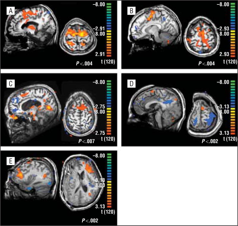
Objectives: To detect covert volitional brain activity in patients with severe brain injury using pattern classification of the blood oxygenation level-dependent (BOLD) response during mental imagery and to compare these results with those of a univariate functional magnetic resonance imaging analysis.
Design: Case-control study.
Setting: Academic research.
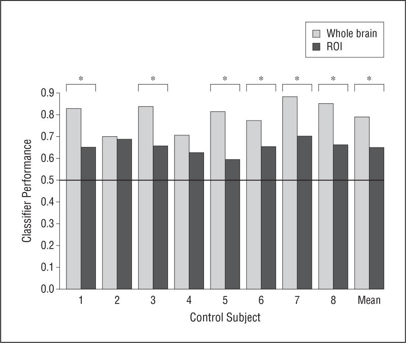
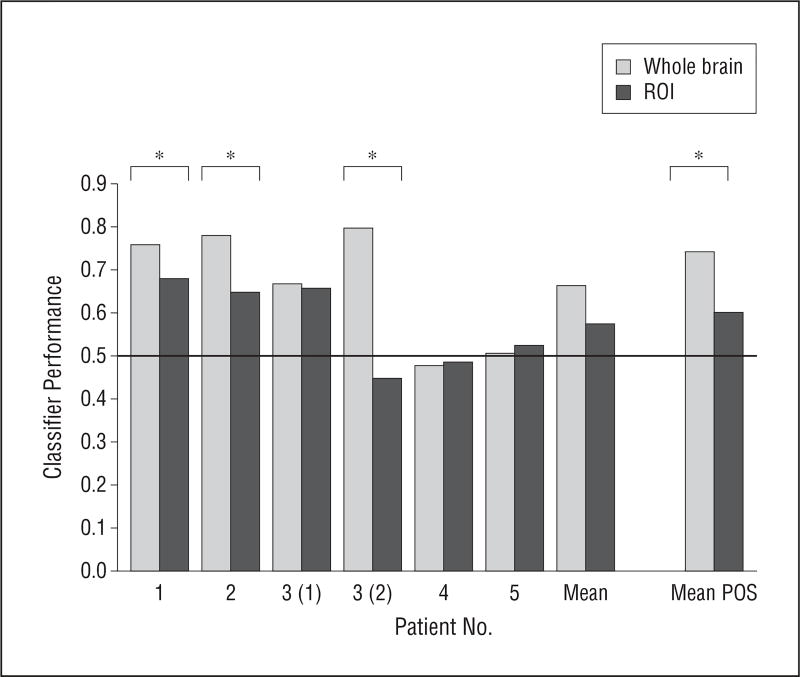
Participants: Experiments were performed in 8 healthy control subjects and in 5 patients with severe brain injury. The patients with severe brain injury constituted a convenience sample.
Main outcome measures: Functional magnetic resonance imaging data were acquired as the patients were asked to follow commands or to answer questions using motor imagery as a proxy response.
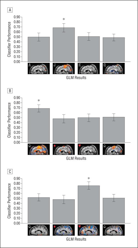
Results: In the controls, the responses were accurately classified. In the patient group, the responses of 3 of 5 patients were correctly classified. The remaining 2 patients showed no significant BOLD response in a standard univariate analysis, suggesting that they did not perform the task. In addition, we showed that a classifier trained on command-following data can be used to evaluate a later communication run. This technique was used to successfully disambiguate 2 potential BOLD responses to a single question.
Conclusions: Pattern classification in functional magnetic resonance imaging is a promising technique for advancing the understanding of volitional brain responses in patients with severe brain injury and may serve as a powerful complement to traditional general linear model-based univariate analysis methods.
Figures
Comment in
-
Wait, wait . . . Don't tell me: tuning in the injured brain.Arch Neurol. 2012 Feb;69(2):158-60. doi: 10.1001/archneurol.2011.1211. Arch Neurol. 2012. PMID: 22332185 No abstract available.
References
-
- Giacino JT, Smart CM. Recent advances in behavioral assessment of individuals with disorders of consciousness. Curr Opin Neurol. 2007;20(6):614–619. - PubMed
-
- Schnakers C, Perrin F, Schabus M, et al. Voluntary brain processing in disorders of consciousness. Neurology. 2008;71(20):1614–1620. - PubMed
-
- Gill-Thwaites H. Lotteries, loopholes and luck: misdiagnosis in the vegetative state patient. Brain Inj. 2006;20(13–14):1321–1328. - PubMed
-
- Monti MM, Vanhaudenhuyse A, Coleman MR, et al. Willful modulation of brain activity in disorders of consciousness. N Engl J Med. 2010;362(7):579–589. - PubMed
Publication types
MeSH terms
Substances
Grants and funding
LinkOut - more resources
Full Text Sources
Medical
