. 2012 May 1;29(7):1518-29.
doi: 10.1089/neu.2011.2030. Epub 2012 Mar 29.

Magnesium sulfate and nimesulide have synergistic effects on rescuing brain damage after transient focal ischemia

Affiliations

Magnesium sulfate and nimesulide have synergistic effects on rescuing brain damage after transient focal ischemia

Liang-Chao Wang et al. J Neurotrauma. .

Abstract

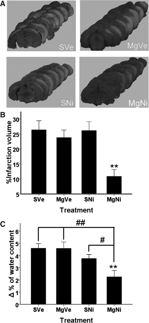
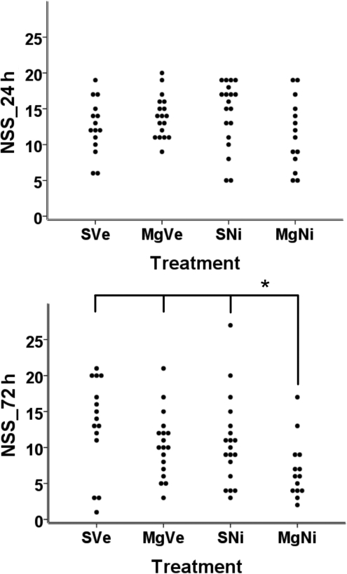
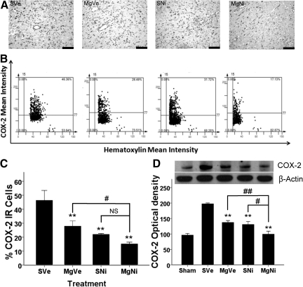
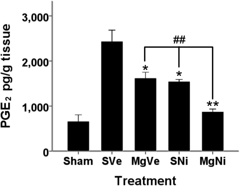
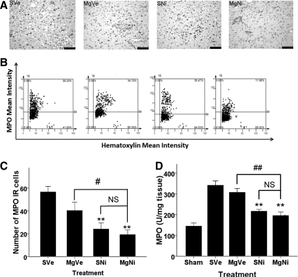
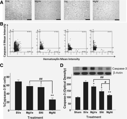
Magnesium sulfate and nimesulide are commonly used drugs with reported neuroprotective effects. Their combination as stroke treatment has the potential benefits of decreasing individual drug dosage and fewer adverse effects. This study evaluated their synergistic effects and compared a low-dose combination with individual drug alone and placebo. Sprague-Dawley rats underwent 90 min of focal ischemia with intraluminal suture occlusion of the middle cerebral artery followed by reperfusion. The rats were randomly assigned to receive one of the following treatments: placebo, magnesium sulfate (MgSO₄; 45 mg/kg) intravenously immediately after the induction of middle cerebral artery occlusion, nimesulide (6 mg/kg) intraperitoneally before reperfusion, and combined therapy. Three days after the ischemia-reperfusion insult, therapeutic outcome was assessed by 2,3,5-triphenyltetrazolium chloride staining and a 28-point neurological severity scoring system. Cyclooxygenase-2, prostaglandin E₂, myeloperoxidase, and caspase-3 expression after treatment were evaluated using Western blot analyses and immunohistochemical staining, followed by immunoreactive cell analysis using tissue cytometry. Only the combination treatment group showed a significant decrease in infarction volume (10.93±6.54% versus 26.43±7.08%, p<0.01), and neurological severity score (p<0.05). Low-dose MgSO₄ or nimesulide showed no significant neuroprotection. There was also significant suppression of cyclooxygenase-2, prostaglandin E₂, myeloperoxidase, and caspase-3 expression in the combination treatment group, suggesting that the combination of the two drugs improved the neuroprotective effects of each individual drug. MgSO₄ and nimesulide have synergistic effects on ischemia-reperfusion insults. Their combination helps decrease drug dosage and adverse effects. Combined treatment strategies may help to combat stroke-induced brain damage in the future.


Figures

FIG. 1.
FIG. 1.
Effect of combined MgSO4 and nimesulide on infarct volume and brain edema. (A) Representative pictures of TTC staining. (B) Percentage of infarction volume shown as mean±standard error of the mean (SEM; n=7–8). Combined treatment with MgSO4 and nimesulide significantly attenuated total infarction volume (**p<0.01 by Dunnett's test), whereas MgSO4 or nimesulide alone had no statistically significant effect on attenuating infarct volume. (C) Brain edema was evaluated by wet-dry weight (n=5–6). Differences in water content between the ipsilateral and contralateral hemispheres (Δ% of water content) 48 h after ischemia-reperfusion injury are represented as mean±SEM. Only combination treatment significantly attenuated brain edema (p=0.002 by Dunnett's test; **p<0.01). Brain edema in the combination treatment group was significantly reduced compared to the groups treated with MgSO4 alone and nimesulide alone (p=0.001 and p=0.024, respectively, by Fisher's protected least-significant difference test; #p<0.05; ##p<0.01; MgSO4, magnesium sulfate; TTC, 2,3,5-triphenyltetrazolium chloride; PVP, polyvinylpyrrolidone; group SVe, treated with placebo [0.25 mL normal saline and 0.25 mL 2% PVP]; group MgVe, treated with MgSO4 45 mg/kg and 0.25 mL 2% PVP; group SNi, treated with 0.25 mL normal saline and nimesulide 6 mg/kg; and group MgNi, treated with MgSO4 45 mg/kg and nimesulide 6 mg/kg).
FIG. 2.
FIG. 2.
Effect of combined treatment on functional outcome after MCAO with reperfusion. Scattergrams of Neurological Severity Score (NSS) per treatment group are shown (n=15–20). (A) There were no significant differences in NSS for all groups 24 h after MCAO. (B) Combined treatment significantly reduced the neurologic deficit seen after MCAO with 72 h reperfusion compared to groups treated with vehicle, MgSO4 only, and nimesulide only (p=0.015, 0.04., and 0.021, respectively, by; Mann-Whitney U test; MCAO, middle cerebral artery occlusion; MgSO4, magnesium sulfate; PVP, polyvinylpyrrolidone; group SVe, treated with placebo [0.25 mL normal saline and 0.25 mL 2% PVP]; group MgVe, treated with MgSO4 45 mg/kg and 0.25 mL 2% PVP; group SNi, treated with 0.25 mL normal saline and nimesulide 6 mg/kg; and group MgNi, treated with MgSO4 45 mg/kg and nimesulide 6 mg/kg).
FIG. 3.
FIG. 3.
Effect of combined MgSO4 and nimesulide on COX-2 expression 72 h after ischemia-reperfusion injury. (A) Representative images of immunohistochemistry for COX-2 (scale bars=100 μm). (B) Representative images of tissue cytometry using TissueQuest software. COX-2-immunoreactive (IR) positive and negative cells were counted and the signal intensity was quantified. (C) The amount of COX-2 IR cells in the inner boundary zone of infarction was recorded as the percentage of IR cells in all cells (n=5 per group). Data are presented as mean±standard error of the mean (SEM). All treatments suppressed COX-2 protein expression compared to the placebo group (p<0.001 for MgNi; p=0.007 for SNi; p=0.021 for MgVe; **p<0.01 by Dunnett's test). Combined treatment improved the effect of MgSO4 (p=0.02 by least-significant difference [LSD] test). The difference between groups receiving nimesulide alone or combined treatment was not statistically significant (p=0.163; #p<0.05; NS, not significant). (D) Western blot analysis of brain tissue for COX-2 protein 72 h after ischemia-reperfusion injury. Single or combined treatments significantly reduced COX-2 protein expression 72 h after MCAO with reperfusion (n=4–5; **p<0.001 by Dunnett's test). Data are presented as mean±SEM. Combined treatment showed improved effects on reducing COX-2 expression compared to treatment with MgSO4 or nimesulide alone (p=0.004 and 0.011, respectively, by LSD test; #p<0.05; ##p<0.01; group SVe, treated with placebo [0.25 mL normal saline and 0.25 mL 2% PVP]; group MgVe, treated with MgSO4 45 mg/kg and 0.25 mL 2% PVP; group SNi, treated with 0.25 mL normal saline and nimesulide 6 mg/kg; and group MgNi, treated with MgSO4 45 mg/kg and nimesulide 6 mg/kg; COX-2, cyclooxygenase-2; MgSO4, magnesium sulfate; PVP, polyvinylpyrrolidone).
FIG. 4.
FIG. 4.
ELISA of PGE2 expression in brain 72 h after ischemic-reperfusion injury. The expression of PGE2 after ischemic-reperfusion injury is shown here (n=4–5). Data are presented as mean±standard error of the mean. PGE2 concentrations in the brain tissue were significantly reduced by both MgSO4 or nimesulide alone (*p<0.01, **p<0.001 by Dunnett's test). Combined treatment significantly reduced the PGE2 concentration compared to the group treated with nimesulide only (##p<0.01; ELISA, enzyme-linked immunosorbent assay; PGE2, prostaglandin E2; MgSO4, magnesium sulfate; PVP, polyvinylpyrrolidone; group SVe, treated with placebo [0.25 mL normal saline and 0.25 mL 2% PVP]; group MgVe, treated with MgSO4 45 mg/kg and 0.25 mL 2% PVP; group SNi, treated with 0.25 mL normal saline and nimesulide 6 mg/kg; and group MgNi, treated with MgSO4 45 mg/kg and nimesulide 6 mg/kg).
FIG. 5.
FIG. 5.
Effect of combined MgSO4 and nimesulide on MPO expression 72 h after ischemia-reperfusion injury. (A) Representative images of immunohistochemistry for MPO (scale bars=100 μm). (B) Representative scatterplots of tissue cytometry using TissueQuest software. MPO immunoreactive-(IR) positive and negative cells were counted, and the signal intensity was quantified. (C) Immunohistochemistry (n=5 per group) showed the number of MPO IR cells in the inner boundary zone of infarction. Data are presented as mean±standard error of the mean. Data showed that MPO IR cells significantly decreased in nimesulide only and combination (p=0.004 and 0.001 respectively Dunnett's test). **p<0.01;. Differences between the combination treatment group and nimesulide only group was not significant, but the difference between the MgSO4 only and the combination treatment groups was significant (#p<0.05, NS: p>0.05). (D) ELISA of MPO expression 72 hours after ischemia-reperfusion. (n=5 per group). MPO concentration in brain tissue is presented as mean±standard error of the mean. MPO expression was not suppressed by treatment with MgSO4 alone. Treatment with nimesulide alone or combined with MgSO4 showed significant suppression compared to the placebo group (**p<0.01 by Dunnett's test), but the difference between these two groups was not significant (##p<0.01 by least-significant difference test; NS, not significant; ELISA, enzyme-linked immunosorbent assay; MgSO4, magnesium sulfate; MPO, myeloperoxidase; PVP, polyvinylpyrrolidone; group SVe, treated with placebo [0.25 mL normal saline and 0.25 mL 2% PVP]; group MgVe, treated with MgSO4 45 mg/kg and 0.25 mL 2% PVP; group SNi, treated with 0.25 mL normal saline and nimesulide 6 mg/kg; and group MgNi, treated with MgSO4 45 mg/kg and nimesulide 6 mg/kg).
FIG. 6.
FIG. 6.
Effect of combined MgSO4 and nimesulide on caspase-3 expression 72 h after ischemia-reperfusion injury. (A) Immunohistochemistry in the inner boundary zone of infarction for caspase-3 (scale bars=100 μm). (B) Representative images of tissue cytometry using TissueQuest software. Caspase-3-immunoreactive (IR) positive and negative cells were counted and the signal intensity was quantified. (C) The amount of caspase-3-IR cells were recorded as percentages of IR cells in all cells, and are presented as mean±standard error of the mean (SEM; n=5 per group). The amount of IR cells in the inner boundary zone of infarction was significantly decreased only in the combination treatment group (**p<0.01 by Dunnett's test). The difference between the nimesulide-only and the combination treatment group was also statistically significant (##p<0.01 by least-significant difference test). (D) Western blot analysis of caspase-3 expression in brain tissue 72 h after ischemia-reperfusion injury (n=5 per group). Data are presented as mean±SEM. Administration of MgSO4 alone or nimesulide alone significantly attenuated caspase-3 expression in brain tissue (*p<0.05, **p<0.01 by Dunnett's test). The combination treatment significantly decreased caspase-3 expression in brain tissue after MCAO with reperfusion (MCAO, middle cerebral artery occlusion; MgSO4 magnesium sulfate; group SVe, treated with placebo [0.25 mL normal saline and 0.25 mL 2% PVP]; group MgVe, treated with MgSO4 45 mg/kg and 0.25 mL 2% PVP; group SNi, treated with 0.25 mL normal saline and nimesulide 6 mg/kg; and group MgNi, treated with MgSO4 45 mg/kg and nimesulide 6 mg/kg; PVP, polyvinylpyrrolidone).

Similar articles

Cited by

References

    1. Allan S.M. Rothwell N.J. Cytokines and acute neurodegeneration. Nat. Rev. Neurosci. 2001;2:734–744. - PubMed
    1. Barone F.C. ad Feuerstein G.Z. Inflammatory mediators and stroke: new opportunities for novel therapeutics. J. Cereb. Blood Flow Metab. 1999;19:819–834. - PubMed
    1. Breckwoldt M.O. Chen J.W. Stangenberg L. Aikawa E. Rodriguez E. Qiu S. Moskowitz M.A. Weissleder R. Tracking the inflammatory response in stroke in vivo by sensing the enzyme myeloperoxidase. Proc. Natl. Acad. Sci. USA. 2008;105:18584–18589. - PMC - PubMed
    1. Candelario-Jalil E. Fiebich B.L. Cyclooxygenase inhibition in ischemic brain injury. Curr. Pharm. Des. 2008;14:1401–1418. - PubMed
    1. Candelario-Jalil E. Gonzalez-Falcon A. Garcia-Cabrera M. Leon O.S. Fiebich B.L. Post-ischaemic treatment with the cyclooxygenase-2 inhibitor nimesulide reduces blood-brain barrier disruption and leukocyte infiltration following transient focal cerebral ischaemia in rats. J. Neurochem. 2007;100:1108–1120. - PubMed

Publication types