. 2012 Mar 5;9(3):514-22.
doi: 10.1021/mp200526m. Epub 2012 Feb 22.

Enhanced tumor treatment using biofunctional indocyanine green-containing nanostructure by intratumoral or intravenous injection

Affiliations

Enhanced tumor treatment using biofunctional indocyanine green-containing nanostructure by intratumoral or intravenous injection

Xiaohui Zheng et al. Mol Pharm. .

Abstract

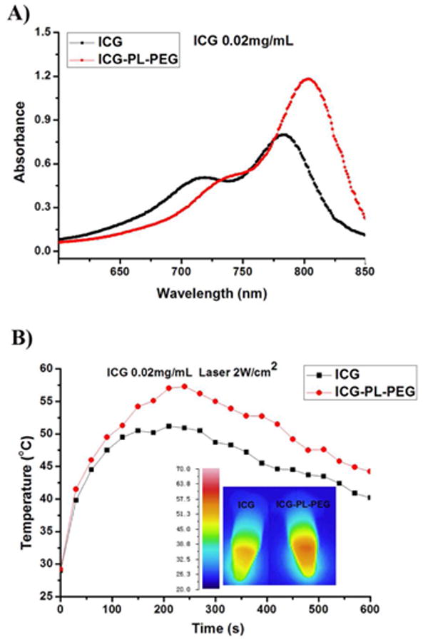
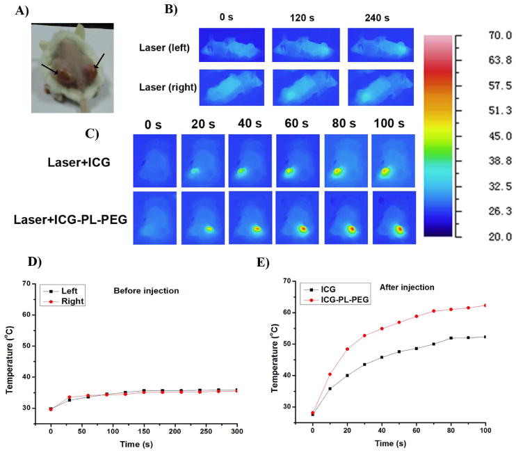
Indocyanine green (ICG) is a conventional dye that can be used in clinical near-infrared (NIR) imaging, and it is also an effective light absorber for laser-mediated photothermal therapy. However, applications of ICG were limited due to its fast degradation in aqueous media and quick clearance from the body. Herein, an ICG-containing nanostructure, ICG-PL-PEG, was developed for photothermal therapy, which was self-assembled by ICG and phospholipid-polyethylene glycol (PL-PEG). Our in vitro and in vivo experiments demonstrated that ICG-PL-PEG suspension was more efficient in producing a NIR-dependent temperature increase than ICG alone, due to the increase of ICG monomers from the addition of PL-PEG to match the central wavelength of the 808 nm laser. When conjugated with integrin α(v)β(3) monoclonal antibody (mAb), ICG-PL-PEG could be selectively internalized and retained in target tumor cells. Irradiation of an 808 nm laser after intravenous administration of ICG-PL-PEG-mAb resulted in tumor suppression in mice, while ICG alone had only limited effect. This is the first time an ICG-containing nanostructure has been used through systemic administration to achieve an efficient in vivo photothermal effect for cancer treatment. Therefore, ICG-PL-PEG could be used as a fluorescent marker as well as a light-absorber for imaging-guided photothermal therapy. All the components of ICG-PL-PEG have been approved for human use. Therefore, this unique ICG-containing nanostructure has great potential in clinical applications.


Figures

Figure 1
Figure 1
Absorption spectra and temperature increases of ICG and ICG-PL-PEG. A. UV-vis-NIR absorption spectra of ICG and ICG-PL-PEG. B. Temperature increases in ICG or ICG-PL-PEG formulation in response to irradiation of an 808-nm laser with a power density of 2 W/cm2 and a duration of 10 minutes. Inset: thermographic images of ICG and ICG-PL-PEG in vials after laser irradiation for 4 min.
Figure 2
Figure 2
Temperature increase in tumor tissue with or without ICG or ICG-PL-PEG in response to irradiation of an 808-nm laser with a power density of 0.5 W/cm2 and different durations. A. Photograph of a mouse bearing bilateral tumors. B and D. Thermographic image and plots of average temperature increase of bilateral tumors during 808-nm laser irradiation without ICG or ICG-PL-PEG. C and E. Thermographic image and plots of average temperature increase of bilateral tumors during 808-nm laser irradiation 3 hours after injection of ICG (left tumor) and ICG-PL-PEG (right tumor).
Figure 3
Figure 3
A. Confocal images of U87-MG cells and MCF-7 cells after incubation in a solution of ICG-PL-PEG-mAb/FITC. U87-MG cells with overexpressed integrin αvβ3 show a high level of uptake of the targeting probe while MCF-7 cells with a low level of integrin αvβ3 show a low level of uptake. The fluorescence emissions from ICG (red) and FITC (green) were co-localized in U87-MG cancer cells, but absent in non-target cells (MCF-7), which was clear evidence that ICG-PL-PEG-mAb/FITC remained stable after entering target cells. B. High-magnification images of fluorescence emission from cells.
Figure 4
Figure 4
In vivo biodistribution analysis of ICG and ICG-PL-PEG-mAb. A. The whole body NIR fluorescent images of mice were obtained six hours after intravenous injection. Detection was performed using the LI-COR Odyssey Infrared Imaging System through 800 nm channel (LI-COR, Inc. Lincoln, NE). ICG was mainly accumulated in liver, while ICG-PL-PEG-mAb had a high accumulation in both tumor and liver. B.U87-MG tumor-bearing mice were sacrificed and major organs were collected for fluorescence imaging. Spectrally resolved ex vivo fluorescence images of different organs were displayed. T: tumor, LI: liver, SP: spleen, LU: lung, K: kidney, H: heart. C. Semiquantitative biodistribution of ICG and ICG-PL-PEG-mAb in mice determined by the averaged fluorescence intensity of each organ (after subtraction by the fluorescence intensity of each organ before injection). P values were calculated using non-paired two-tailed Student's t-test (*P <0.05, **P<0.01, ***P<0.001; n=3 per group).
Figure 5
Figure 5
Photothermal treatment of mouse tumor using intravenous injection of ICG and ICG-PL-PEG-mAb, followed by laser irradiation. A. Photograph of a mouse bearing U87-MG tumor. B. Thermographic images of mice bearing U87-MG tumors under different treatments. C. Plot of maximum surface temperature of the irradiated area as a function of the irradiation time. (n=3 per group). D. Histological staining of the excised tumors 12 hours after different treatments. Distinctive characteristics of cellular damage were observed in the Laser+ICG-PL-PEG-mAb treated tumors, including coagulative necrosis (arrow), abundant pyknosis (arrowhead) and considerable regions of karyolysis (asterisk). E. Time-dependent tumor growth curves of U87-MG tumor. The results were presented as the arithmetic means with standard deviations of tumor volumes in each group. Only the Laser+ICG-PL-PEG-mAb treated group shows significant suppression of tumor growth compared with other experimental groups (n=3).

References

    1. Nolsoe CP, Torp-Pedersen S, Burcharth F, Horn T, Pedersen S, Christensen NE, Olldag ES, Andersen PH, Karstrup S, Lorentzen T, et al. Interstitial hyperthermia of colorectal liver metastases with a US-guided Nd-YAG laser with a diffuser tip: a pilot clinical study. Radiology. 1993;187:333–337. - PubMed
    1. Amin Z, Donald JJ, Masters A, Kant R, Steger AC, Bown SG, Lees WR. Hepatic metastases: interstitial laser photocoagulation with real-time US monitoring and dynamic CT evaluation of treatment. Radiology. 1993;187:339–347. - PubMed
    1. Anghileri LJ, Robert J. Hyperthermia in Cancer Treatment. CRC Press; Boca Raton FL: 1986. pp. 59–78.
    1. Weissleder R. A clearer vision for in vivo imaging. Nat Biotechnol. 2001;19:316–317. - PubMed
    1. Chen J, Glaus C, Laforest R, Zhang Q, Yang M, Gidding M, Welch MJ, Xia Y. Gold Nanocages as Photothermal Transducers for Cancer Treatment. small. 2010;6:811–817. - PMC - PubMed

Publication types