Review
. 2012 Jun;26(2):132-43.
doi: 10.3109/01677063.2011.647143. Epub 2012 Feb 14.

From Drosophila to humans: reflections on the roles of the prolyl isomerases and chaperones, cyclophilins, in cell function and disease

Affiliations
Review

From Drosophila to humans: reflections on the roles of the prolyl isomerases and chaperones, cyclophilins, in cell function and disease

Paulo A Ferreira et al. J Neurogenet. 2012 Jun.

Abstract

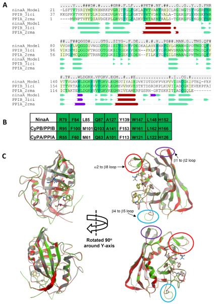
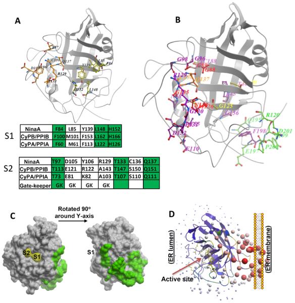
Despite remarkable advances in human genetics and other genetic model systems, the fruit fly, Drosophila melanogaster, remains a powerful experimental tool to probe with ease the inner workings of a myriad of biological and pathological processes, even when evolutionary forces impart apparent divergences to some of such processes. The understanding of such evolutionary differences provides mechanistic insights into genotype-phenotype correlations underpinning biological processes across metazoans. The pioneering work developed by the William Pak laboratory for the past four decades, and the work of others, epitomize the notion of how the Drosophila system breaks new fertile ground or complements research fields of high scientific and medical relevance. Among the three major genetic complementation groups produced by the Pak's laboratory and impairing distinct facets of photoreceptor neuronal function, the nina group (ninaA, …., ninaJ) selectively affects the biogenesis of G protein-coupled receptors (GPCRs), mediating the photoconversion and transduction of light stimuli. Among the nina genes identified, ninaA arguably assumes heightened significance for several reasons. First, it presents unique physiological selectivity toward the biogenesis of a subset of GPCRs, a standalone biological manifestation yet to be discerned for most mammalian homologues of NinaA. Second, NinaA belongs to a family of proteins, immunophilins, which are the primary targets for immunosuppressive drugs at the therapeutic forefront of a multitude of medical conditions. Third, NinaA closest homologue, cyclophilin B (CyPB/PPIB), is an immunophilin whose loss-of-function was found recently to cause osteogenesis imperfecta in the human. This report highlights advances made by studies on some members of immunophilins, the cyclophilins. Finally, it reexamines critically data and dogmas derived from past and recent genetic, structural, biological, and pathological studies on NinaA and few other cyclophilins that support some of such paradigms to be less than definite and advance our understanding of the roles of cyclophilins in cell function, disease, and therapeutic interventions.


Figures

FIGURE 1
FIGURE 1
Molecular modeling of NinaA to CyPB/PPIB and CyPA/PPIA. (A) Sequence alignment of NinaA, CyPB/PPIB, CyPA/PPIA. Identical and conserved residues are highlighted in green and yellow, respectively. NinaA exhibits 49% and 43% identity to CyPB/PPIB and CyPA/PPIA, respectively. Consensus sequence is noted above the alignment. Consensus of secondary-structure elements are shown below the primary sequence alignment. The secondary structure assignment is taken from the PDB files 2rma and 3ici for CyPA/PPIA and CyPB/PPIB, respectively, and the model of NinaA. Legend: Green arrows and solid green triangles, β-sheet; red cylinders and spheres, α-helices; purple cylinders, non-canonical helices. (B) Conservation of residues comprising the PPIase active site of NinaA, CyPB/PPIB and CyPA/PPIA. Identical residues are highlighted in green. (C) Structural superposition of the NinaA model (green ribbon), the CyPB/PPIB template crystal structure (red ribbon), and the CyPA/PPIA crystal structure (grey ribbon). The residues of the PPIase active site are shown in stick representation and the carbon atoms are colored red and orange, respectively. The main structural differences between the three structures are in three loop regions: β1 to β2 (circled in purple), β4 to β5 (circled in light blue), and β2 to β8 (circled in red). The β1 to β2 loop turn is conserved between the NinaA model and the PPIB template and this turn differs from the turn seen in PPIA structures. A two residue insertion in the β4 to β5 loop in NinaA gives rise to a different loop structure in this region compared to PPIA and PPIB. The α2 to β8 loop is one residue shorter in PPIA and therefore differs to the loops seen in NinaA and PPIB. The modeling indicates that there may be an additional β-sheet in this loop in NinaA compared to PPIA and PPIB.
Figure 2
Figure 2
Assignment of physiological relevant residues of NinaA to new cylophilin domains. (A) Location of residues comprising S1 and S2 pockets as defined by Davis et al, 2010, in NinaA 3D-structure. Conservation of residues comprising the S1 and S2 pockets of NinaA, CyPB/PPIB and CyPA/PPIA are shown in the tables below the 3D-structure. The conservation of gatekeeper residues is also depicted. Identical residues are highlighted in green. (B) Mapping of residues caused by mutations in ninaA (Ondek et al., 1992; Table 1) to the 3D-model structure of NinaA. The mutations were grouped in five groups based on their location. Residues nearby the S1 pocket are in yellow, residues in the S2 pocket are in orange, residues in the S2 extended (S2e) region are in red, residues comprising Pm domain facing the membrane surface are in green, all other residues are in magenta. Note F198 points away from the surface. The S2e region comprises the residues, G88, G89, M126, N128 and G135. All residues, but N128, are buried in a shell next to the periphery of S2 binding pocket. (C) Surface representation of residues of Pm domain of NinaA and their relative location to S1 and S2 pockets. (D) Ribbon representation of the relative orientation of the PPIase active site of NinaA to its Pm domain. Active site residues are represented in the purple ribbons by sticks in yellow.
Figure 3
Figure 3
Docking of Rh1, Rh2, Rh3 and Rh4 prolyl-peptide substrates of opsins of Drosophila and equivalent to P23 of human rhodopsin to S1 and S2 pockets of NinaA. (A) Alignment of Rh1, Rh2, Rh3 and Rh4 prolyl-peptide sequences of opsins of Drosophila comprising the equivalent P23 (in bold) of human rhodopsin. Only Rh1 and Rh2 scored as binders according to their fit in the S1 and S2 pockets of NinaA and with the Rh1 being a better binder. (B, C) are surface representations of NinaA upon docking of Rh1 (B) and Rh2 (C) prolyl substrates shown in (A) to its S1 and S2 pockets.

References

    1. Abagyan R, Orry A, Raush E, Totrov M. ICM User Guide 3.7. La Jolla, CA; Molsoft LLC: 2010.
    1. Abagyan R, Totrov M. Biased probability Monte Carlo conformational searches and electrostatic calculations for peptides and proteins. J Mol Biol. 1994;235:983–1002. - PubMed
    1. Abagyan R, Totrov M, Kuznetsov D. ICM-a new method for protein modeling and design: applications to docking and structure prediction from the distorted native conformation. J Comput Chem. 1994;15:488–506.
    1. Abagyan RA, Batalov S. Do aligned sequences share the same fold? J Mol Biol. 1997;273:355–368. - PubMed
    1. Allain F, Vanpouille C, Carpentier M, Slomianny MC, Durieux S, Spik G. Interaction with glycosaminoglycans is required for cyclophilin B to trigger integrin-mediated adhesion of peripheral blood T lymphocytes to extracellular matrix. Proceedings of the National Academy of Sciences of the United States of America. 2002;99:2714–2719. - PMC - PubMed

LinkOut - more resources