

Diet supplementation with green tea extract epigallocatechin gallate prevents progression to glucose intolerance in db/db mice
- PMID: 22333133
- PMCID: PMC3298777
- DOI: 10.1186/1743-7075-9-11
Diet supplementation with green tea extract epigallocatechin gallate prevents progression to glucose intolerance in db/db mice
Abstract
Background: Green tea was suggested as a therapeutic agent for the treatment of diabetes more than 70 years ago, but the mechanisms behind its antidiabetic effect remains elusive. In this work, we address this issue by feeding a green tea extract (TEAVIGO™) with a high content of epigallocatechin gallate (EGCG) or the thiazolidinedione PPAR-γ agonist rosiglitazone, as positive control, to db/db mice, an animal model for diabetes.
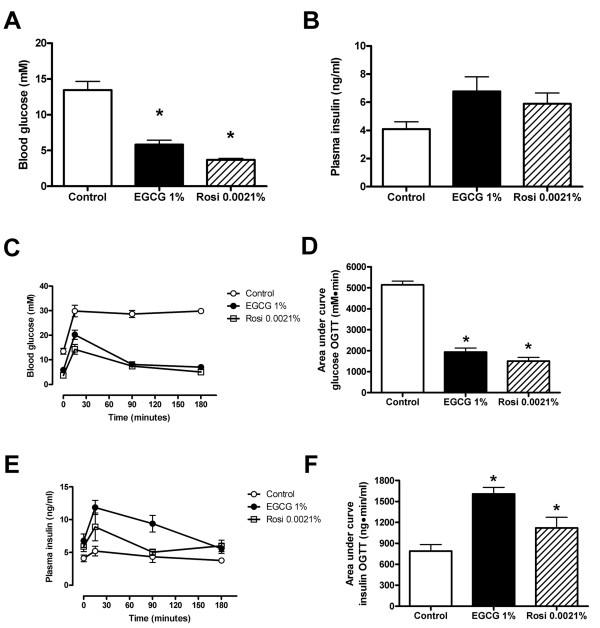
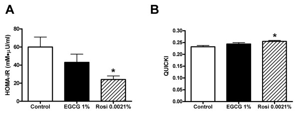
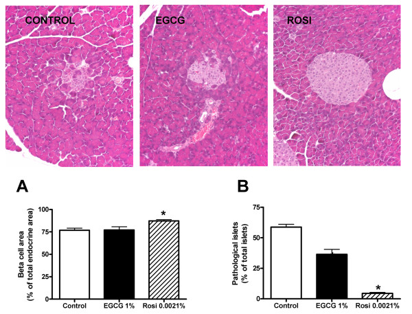
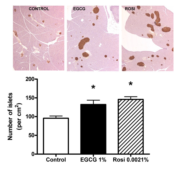
Methods: Young (7 week-old) db/db mice were randomized and assigned to receive diets supplemented with or without EGCG or rosiglitazone for 10 weeks. Fasting blood glucose, body weight and food intake was measured along the treatment. Glucose and insulin levels were determined during an oral glucose tolerance test after 10 weeks of treatment. Pancreata were sampled at the end of the study for blinded histomorphometric analysis. Islets were isolated and their mRNA expression analyzed by quantitative RT-PCR.
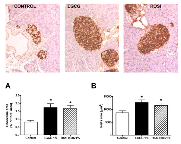
Results: The results show that, in db/db mice, EGCG improves glucose tolerance and increases glucose-stimulated insulin secretion. EGCG supplementation reduces the number of pathologically changed islets of Langerhans, increases the number and the size of islets, and heightens pancreatic endocrine area. These effects occurred in parallel with a reduction in islet endoplasmic reticulum stress markers, possibly linked to the antioxidative capacity of EGCG.
Conclusions: This study shows that the green tea extract EGCG markedly preserves islet structure and enhances glucose tolerance in genetically diabetic mice. Dietary supplementation with EGCG could potentially contribute to nutritional strategies for the prevention and treatment of type 2 diabetes.
Figures
Similar articles
-
Epigallocatechin gallate supplementation alleviates diabetes in rodents.J Nutr. 2006 Oct;136(10):2512-8. doi: 10.1093/jn/136.10.2512. J Nutr. 2006. PMID: 16988119
-
Decaffeinated green tea extract rich in epigallocatechin-3-gallate prevents fatty liver disease by increased activities of mitochondrial respiratory chain complexes in diet-induced obesity mice.J Nutr Biochem. 2015 Nov;26(11):1348-56. doi: 10.1016/j.jnutbio.2015.07.002. Epub 2015 Jul 26. J Nutr Biochem. 2015. PMID: 26300331
-
Epigallocatechin gallate reduces vascular inflammation in db/db mice possibly through an NF-κB-mediated mechanism.Mol Nutr Food Res. 2012 Sep;56(9):1424-32. doi: 10.1002/mnfr.201200040. Epub 2012 Jul 2. Mol Nutr Food Res. 2012. PMID: 22753231 Free PMC article.
-
Dietary Epigallocatechin-3-Gallate Alters the Gut Microbiota of Obese Diabetic db/db Mice: Lactobacillus Is a Putative Target.J Med Food. 2020 Oct;23(10):1033-1042. doi: 10.1089/jmf.2020.4700. J Med Food. 2020. PMID: 33054538
-
Green Tea and Epigallocatechin Gallate (EGCG) for the Management of Nonalcoholic Fatty Liver Diseases (NAFLD): Insights into the Role of Oxidative Stress and Antioxidant Mechanism.Antioxidants (Basel). 2021 Jul 5;10(7):1076. doi: 10.3390/antiox10071076. Antioxidants (Basel). 2021. PMID: 34356308 Free PMC article. Review.
Cited by
-
Can Tea Consumption be a Safe and Effective Therapy Against Diabetes Mellitus-Induced Neurodegeneration?Curr Neuropharmacol. 2014 Dec;12(6):475-89. doi: 10.2174/1570159X13666141204220539. Curr Neuropharmacol. 2014. PMID: 25977676 Free PMC article.
-
Synergistic Hypolipidemic Effects and Mechanisms of Phytochemicals: A Review.Foods. 2022 Sep 9;11(18):2774. doi: 10.3390/foods11182774. Foods. 2022. PMID: 36140902 Free PMC article. Review.
-
Regeneration of Pancreatic β-Cells for Diabetes Therapeutics by Natural DYRK1A Inhibitors.Metabolites. 2022 Dec 29;13(1):51. doi: 10.3390/metabo13010051. Metabolites. 2022. PMID: 36676976 Free PMC article. Review.
-
Green tea consumption and risk of type 2 diabetes in Chinese adults: the Shanghai Women's Health Study and the Shanghai Men's Health Study.Int J Epidemiol. 2018 Dec 1;47(6):1887-1896. doi: 10.1093/ije/dyy173. Int J Epidemiol. 2018. PMID: 30169796 Free PMC article.
-
Role of PCK1 gene on oil tea-induced glucose homeostasis and type 2 diabetes: an animal experiment and a case-control study.Nutr Metab (Lond). 2019 Feb 13;16:12. doi: 10.1186/s12986-019-0337-8. eCollection 2019. Nutr Metab (Lond). 2019. PMID: 30805021 Free PMC article.
References
-
- Katiyar SK, Mukhtar H. Tea consumption and cancer. World Rev Nutr Diet. 1996;79:154–184. - PubMed
LinkOut - more resources
Full Text Sources
Miscellaneous
