Review
. 2012 Apr;1825(2):229-44.
doi: 10.1016/j.bbcan.2012.01.004. Epub 2012 Feb 7.

The p53 circuit board

Affiliations
Review

The p53 circuit board

Kelly D Sullivan et al. Biochim Biophys Acta. 2012 Apr.

Abstract

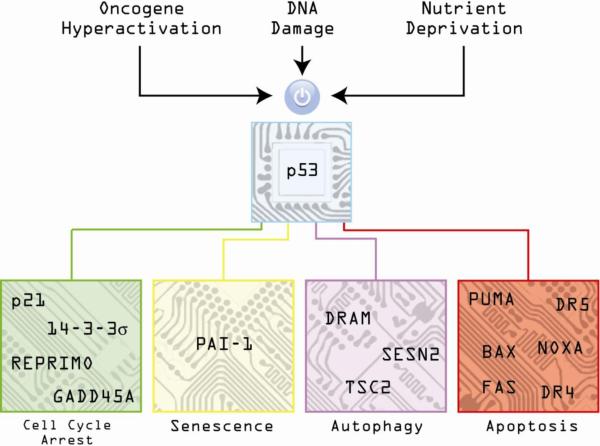
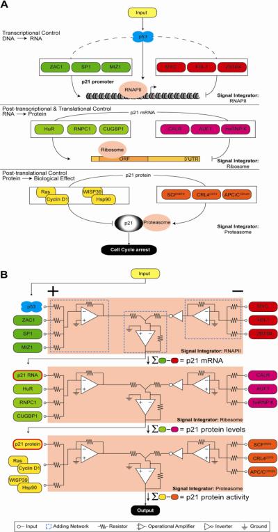
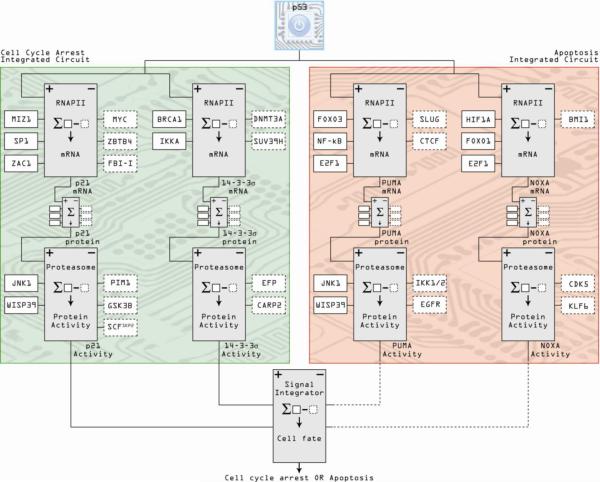
The p53 tumor suppressor is embedded in a large gene network controlling diverse cellular and organismal phenotypes. Multiple signaling pathways converge onto p53 activation, mostly by relieving the inhibitory effects of its repressors, MDM2 and MDM4. In turn, signals originating from increased p53 activity diverge into distinct effector pathways to deliver a specific cellular response to the activating stimuli. Much attention has been devoted to dissecting how the various input pathways trigger p53 activation and how the activity of the p53 protein itself can be modulated by a plethora of co-factors and post-translational modifications. In this review we will focus instead on the multiple configurations of the effector pathways. We will discuss how p53-generated signals are transmitted, amplified, resisted and eventually integrated by downstream gene circuits operating at the transcriptional, post-transcriptional and post-translational levels. We will also discuss how context-dependent variations in these gene circuits define the cellular response to p53 activation and how they may impact the clinical efficacy of p53-based targeted therapies.


Figures

Figure 1
Figure 1. The p53 circuit board
Activation of p53 by diverse stimuli such as oncogene hyperactivation, DNA damage and nutrient deprivation results in increased expression of numerous genes controlling different cellular outcomes such as cell cycle arrest, senescence, autophagy and apoptosis.
Figure 2
Figure 2. The p21 circuit
The p53 target gene p21 is co-regulated by various factors acting at the transcriptional, post-transcriptional and post-translational levels, all of which can potentially affect p53/p21-dependent cell cycle arrest. In both A and B, from top to bottom, transcriptional control of p21 gene expression merges the action of positive and negative transcriptional regulators into a single output, functional mRNA. In this context, p53 is just one among many regulators feeding into RNA polymerase II (RNAPII), the signal integrator at this step, which synthesizes p21 mRNA, the input for the next level of regulation. From there, signals from factors that positively and negatively influence RNA stability and translation are combined via the ribosome signal integrator to achieve protein synthesis. Finally, the levels of active p21 protein in the cell are fine-tuned by stabilizing and destabilizing signals resolved by the proteasome. In A, the regulatory layers are displayed as cartoons of DNA, RNA and protein molecules. In B, the same processes are depicted as an electrical circuit comprising three signal integrators. At the transcriptional level, activating signals including p53 (green) are applied to the positive terminals of the RNAPII signal integrator (+) and are summed internally by an adding network (dashed blue box); inhibitory signals (red, negative terminals) are combined similarly. The difference in total activating and total inhibitory signals determines output amplitude, in this case p21 mRNA level. Integrator output can subsequently serve as input to other signal processing machinery; in this example, p21 mRNA levels become positive inputs for p21 protein production in the ribosome.
Figure 3
Figure 3. Integrated circuits of cell cycle arrest and apoptosis comprise a minimal p53 circuit board
Following a similar schema to Figure 2, co-regulators of individual p53-target genes acting at the transcriptional, post-transcriptional and post-translational levels are integrated into circuits. Individual genes contributing to a particular cellular outcome following p53 activation are then combined into integrated circuits, represented here by the cell cycle arrest and apoptosis integrated circuits composed of p21/14-3-3σ and PUMA/NOXA, respectively. The two integrated circuits are then assembled into the p53 circuit board, which ultimately consolidates all positive and negative signals to define one cell fate. Positive regulators of gene activity are denoted by solid boxes, dashed boxes indicate negative regulators.
Figure 4
Figure 4. The p53 family of transcription factors
Schematic of the gene architecture and well characterized isoforms of p53 and the closely related factors p63 and p73. Arrows represent alternative promoters, boxes represent exons (black segments are untranslated regions). TAD: transactivaiton domain. TA2: second transactivation domain. PY: proline rich domain. DBD: DNA binding domain. NLS: nuclear localization signal. OD: oligomerization domain. SAM: sterile-alpha motif. TID: trans-inhibitory domain.
Figure 5
Figure 5. The impact of p63 and p73 isoforms on the p53 circuit board
This wiring diagram illustrates the ability of various p53 family members to co-regulate, both positively and negatively, canonical p53 target genes involved in cell cycle arrest and apoptosis. Different isoforms of p63 and p73 are expressed in diverse cell types, thus providing cell type-specific regulatory capacity. Furthermore, p63 and p73 isoforms are positively and negatively regulated by diverse factors that do not affect p53 directly (gray boxes on top), thus providing additional opportunities for stimulus-specific regulation within the circuit board.
Figure 6
Figure 6. Context-dependent configurations of the p53 circuit board define the efficacy of p53 based therapies
A. An example of cell type-specific p53 responses is provided by non-genotoxic p53 activation by Nutlin-3 across cancer cell lines. In BV173 cells (left), the cell cycle arrest integrated circuit is impaired by p21 mRNA decay, 14-3-3σ promoter methylation and impaired processing of miR-34a. In contrast, these three cell cycle arrest genes are effectively activated in HCT116 cells (right), where they function coordinately to establish a cell cycle arrest response, even though potent apoptotic genes such as PUMA have also been induced. B. Stimulus-specific assembly of the p53 circuit in response to p53 activation by Nutlin-3 versus 5-FU in HCT116 cells. Both Nutlin-3 and 5-FU strongly activate genes involved in both cell cycle arrest and apoptosis; however, only 5-FU treatment results in p53-independent stabilization of DR4 mRNA and concomitant upregulation of DR4 protein levels, which is required for caspase 8 activation and proteolytic activation of BID into tBID. Activation of the DR4:tBID axis by 5-FU drives the apoptotic response by promoting oligomerization of poised BAX at the mitochondria.

References

    1. DeLeo AB, Jay G, Appella E, Dubois GC, Law LW, Old LJ. Detection of a transformation-related antigen in chemically induced sarcomas and other transformed cells of the mouse. Proc Natl Acad Sci U S A. 1979;76:2420–2424. - PMC - PubMed
    1. Linzer DI, Levine AJ. Characterization of a 54K dalton cellular SV40 tumor antigen present in SV40-transformed cells and uninfected embryonal carcinoma cells. Cell. 1979;17:43–52. - PubMed
    1. Lane DP, Crawford LV. T antigen is bound to a host protein in SV40-transformed cells. Nature. 1979;278:261–263. - PubMed
    1. Eliyahu D, Raz A, Gruss P, Givol D, Oren M. Participation of p53 cellular tumour antigen in transformation of normal embryonic cells. Nature. 1984;312:646–649. - PubMed
    1. Jenkins JR, Rudge K, Currie GA. Cellular immortalization by a cDNA clone encoding the transformation-associated phosphoprotein p53. Nature. 1984;312:651–654. - PubMed

Substances