

The road to functional imaging and ultrahigh fields
- PMID: 22333670
- PMCID: PMC3531996
- DOI: 10.1016/j.neuroimage.2012.01.134
The road to functional imaging and ultrahigh fields
Abstract
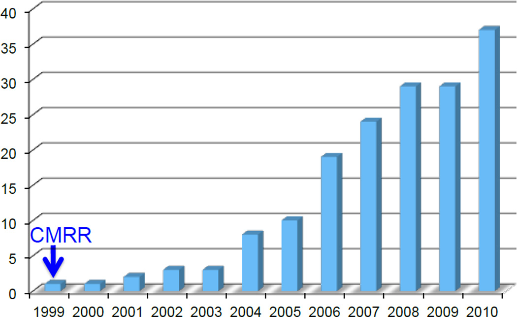
The Center for Magnetic Resonance (CMRR) at the University of Minnesota was one of the laboratories where the work that simultaneously and independently introduced functional magnetic resonance imaging (fMRI) of human brain activity was carried out. However, unlike other laboratories pursuing fMRI at the time, our work was performed at 4T magnetic field and coincided with the effort to push human magnetic resonance imaging to field strength significantly beyond 1.5T which was the high-end standard of the time. The human fMRI experiments performed in CMRR were planned between two colleagues who had known each other and had worked together previously in Bell Laboratories, namely Seiji Ogawa and myself, immediately after the Blood Oxygenation Level Dependent (BOLD) contrast was developed by Seiji. We were waiting for our first human system, a 4T system, to arrive in order to attempt at imaging brain activity in the human brain and these were the first experiments we performed on the 4T instrument in CMRR when it became marginally operational. This was a prelude to a subsequent systematic push we initiated for exploiting higher magnetic fields to improve the accuracy and sensitivity of fMRI maps, first going to 9.4T for animal model studies and subsequently developing a 7T human system for the first time. Steady improvements in high field instrumentation and ever expanding armamentarium of image acquisition and engineering solutions to challenges posed by ultrahigh fields have brought fMRI to submillimeter resolution in the whole brain at 7T, the scale necessary to reach cortical columns and laminar differentiation in the whole brain. The solutions that emerged in response to technological challenges posed by 7T also propagated and continues to propagate to lower field clinical systems, a major advantage of the ultrahigh fields effort that is underappreciated. Further improvements at 7T are inevitable. Further translation of these improvements to lower field clinical systems to achieve new capabilities and to magnetic fields significantly higher than 7T to enable human imaging is inescapable.
Copyright © 2012 Elsevier Inc. All rights reserved.
Figures

Similar articles
-
Development of functional imaging in the human brain (fMRI); the University of Minnesota experience.Neuroimage. 2012 Aug 15;62(2):613-9. doi: 10.1016/j.neuroimage.2012.01.135. Epub 2012 Feb 8. Neuroimage. 2012. PMID: 22342875 Free PMC article. Review.
-
Revealing human ocular dominance columns using high-resolution functional magnetic resonance imaging.Neuroimage. 2012 Aug 15;62(2):1029-34. doi: 10.1016/j.neuroimage.2011.08.086. Epub 2011 Sep 2. Neuroimage. 2012. PMID: 21914484 Review.
-
Ultra high resolution fMRI at ultra-high field.Neuroimage. 2012 Aug 15;62(2):1024-8. doi: 10.1016/j.neuroimage.2012.01.018. Epub 2012 Jan 9. Neuroimage. 2012. PMID: 22245344 Free PMC article. Review.
-
Spin-echo fMRI: The poor relation?Neuroimage. 2012 Aug 15;62(2):1109-15. doi: 10.1016/j.neuroimage.2012.01.003. Epub 2012 Jan 8. Neuroimage. 2012. PMID: 22245351 Review.
-
The intravascular susceptibility effect and the underlying physiology of fMRI.Neuroimage. 2012 Aug 15;62(2):995-9. doi: 10.1016/j.neuroimage.2012.01.113. Epub 2012 Jan 28. Neuroimage. 2012. PMID: 22305989 Review.
Cited by
-
Frequency specific interactions of MEG resting state activity within and across brain networks as revealed by the multivariate interaction measure.Neuroimage. 2013 Oct 1;79:172-83. doi: 10.1016/j.neuroimage.2013.04.062. Epub 2013 Apr 28. Neuroimage. 2013. PMID: 23631996 Free PMC article.
-
Cerebrospinal fluid-suppressed T2 -weighted MR imaging at 7 T for human brain.Magn Reson Med. 2019 May;81(5):2924-2936. doi: 10.1002/mrm.27598. Epub 2018 Nov 19. Magn Reson Med. 2019. PMID: 30450583 Free PMC article.
-
Functional MRI of murine olfactory bulbs at 15.2T reveals characteristic activation patters when stimulated by different odors.Sci Rep. 2023 Aug 16;13(1):13343. doi: 10.1038/s41598-023-39650-0. Sci Rep. 2023. PMID: 37587261 Free PMC article.
-
Advantages of cortical surface reconstruction using submillimeter 7 T MEMPRAGE.Neuroimage. 2018 Jan 15;165:11-26. doi: 10.1016/j.neuroimage.2017.09.060. Epub 2017 Sep 29. Neuroimage. 2018. PMID: 28970143 Free PMC article.
-
Pushing spatial and temporal resolution for functional and diffusion MRI in the Human Connectome Project.Neuroimage. 2013 Oct 15;80:80-104. doi: 10.1016/j.neuroimage.2013.05.012. Epub 2013 May 21. Neuroimage. 2013. PMID: 23702417 Free PMC article.
References
-
- Abduljalil AM, Kangarlu A, Zhang X, Burgess RE, Robitaille PM. Acquisition of human multislice MR images at 8 Tesla. J Comput Assist Tomogr. 1999;23(3):335–340. - PubMed
-
- Adriany G, Van de Moortele PF, Wiesinger F, Moeller S, Strupp JP, Andersen P, Snyder C, Zhang X, Chen W, Pruessmann KP, Boesiger P, Vaughan T, Ugurbil K. Transmit and receive transmission line arrays for 7 Tesla parallel imaging. Magn Reson Med. 2005;53(2):434–445. - PubMed
-
- Atkinson IC, Thulborn KR. Feasibility of mapping the tissue mass corrected bioscale of cerebral metabolic rate of oxygen consumption using 17-oxygen and 23-sodium MR imaging in a human brain at 9.4 T. Neuroimage. 2010;51(2):723–733. - PubMed
-
- Barfuss H, Fischer H, Hentschel D, Ladebeck R, Oppelt A, Wittig R, Duerr W, Oppelt R. In vivo magnetic resonance imaging and spectroscopy of humans with a 4 T whole-body magnet. NMR Biomed. 1990;3(1):31–45. - PubMed
Publication types
MeSH terms
Grants and funding
LinkOut - more resources
Full Text Sources
Medical
