. 2012 Aug;20(8):844-51.
doi: 10.1038/ejhg.2012.20. Epub 2012 Feb 15.

COL4A2 mutation associated with familial porencephaly and small-vessel disease

Affiliations

COL4A2 mutation associated with familial porencephaly and small-vessel disease

Elly Verbeek et al. Eur J Hum Genet. 2012 Aug.

Abstract

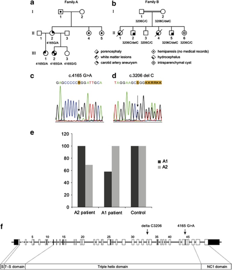
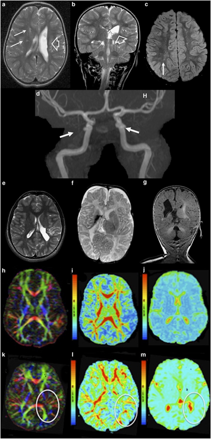
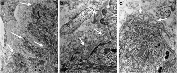
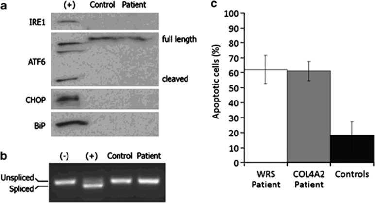
Familial porencephaly, leukoencephalopathy and small-vessel disease belong to the spectrum of disorders ascribed to dominant mutations in the gene encoding for type IV collagen alpha-1 (COL4A1). Mice harbouring mutations in either Col4a1 or Col4a2 suffer from porencephaly, hydrocephalus, cerebral and ocular bleeding and developmental defects. We observed porencephaly and white matter lesions in members from two families that lack COL4A1 mutations. We hypothesized that COL4A2 mutations confer genetic predisposition to porencephaly, therefore we sequenced COL4A2 in the family members and characterized clinical, neuroradiological and biochemical phenotypes. Genomic sequencing of COL4A2 identified the heterozygous missense G1389R in exon 44 in one family and the c.3206delC change in exon 34 leading to frame shift and premature stop, in the second family. Fragmentation and duplication of epidermal basement membranes were observed by electron microscopy in a c.3206delC patient skin biopsy, consistent with abnormal collagen IV network. Collagen chain accumulation and endoplasmic reticulum (ER) stress have been proposed as cellular mechanism in COL4A1 mutations. In COL4A2 (3206delC) fibroblasts we detected increased rates of apoptosis and no signs of ER stress. Mutation phenotypes varied, including porencephaly, white matter lesions, cerebellar and optic nerve hypoplasia and unruptured carotid aneurysm. In the second family however, we found evidence for additional factors contributing to the phenotype. We conclude that dominant COL4A2 mutations are a novel major risk factor for familial cerebrovascular disease, including porencephaly and small-vessel disease with reduced penetrance and variable phenotype, which might also be modified by other contributing factors.


Figures

Figure 1
Figure 1
Pedigrees of families A and B. Different symbols indicate individuals affected with cerebral vascular disease, while the genotype is indicated under the symbol. The genotype of the tested individuals is indicated: (a) 4165G/G=wild-type sequence; 4165G/A=heterozygous mutation; (b) 3206C/C=wild-type sequence; 3206C/delC=heterozygous mutation. Electroferograms indicate the heterozygous mutation in the COL4A2 sequence of family A c.4165G>A (p.G1389R) (c) and family B c.3206delC leading to frame shift (d). Quantitative, real time PCR analysis showing relative expression of COL4A1 and COL4A2 mRNA in fibroblasts from patient II.2 (family B) with c.3206delC mutation (A2 patient) and a patient bearing a pathogenic c.3321G>C (p.G1067A) mutation in exon 38 of COL4A1 (A1 patient), compared with a control cell line. Results are average of two separate experiments. For each cell line the highest expression is set as 100% on the Y axis (e). Position of the mutations within the schematic representation of the COL4A2 genomic organization (f).
Figure 2
Figure 2
Brain MRI of patients with COL4A2 mutations. (a, b) Axial and coronal T2 of patient III.2 from family A at the age of 2 years indicate ex-vacuo dilation of the left lateral ventricle (porencephaly) (open arrow) and periventricular white matter lesions (solid arrows) resulting from presumed perinatal stroke. (c) FLAIR image illustrates T2 prolongation in the white matter – in patient III.1 from family A at the age of 8 years, suggesting gliosis (arrow). (d) MR angiography of their mother (subject II.1, family A) at adult age shows bilateral internal carotid aneurysms (at the level of cavernous sinus). (e) Axial T2 weighted images of Patient II.2, family B, at the age of 15 years show a porencephalic dilatation of the left occipital ventricle. (f, g) Axial T2 weighted and coronal FLAIR images of patient II.5 from family B at the age of 5 months show porencephaly of the right ventricle with hypoplastic left cerebellar hemisphere. (hm) Reconstruction of the white matter tracts obtained from magnetic resonance diffusion tensor imaging (MR-DTI) data. (k,l) Patient II-2 (family B) reveals reduced fractional anisotropy in the left radiation optica and tractus corticospinalis at the side of the porencephalic lesion (encircled), compared with an age-matched control (h,i). A restriction of the total ADC was observed in the whole cerebral white matter of the patient (m), compared with an age-matched control (j).
Figure 3
Figure 3
Skin transmission electron microscopy. Upper arm skin biopsy of subject II-2 of family B was analysed by transmission electron microscopy according to Plaisier et al in double blind experiments by two investigators and independently scored. (a) Control skin biopsy showing illustrative digitations (arrows) of the epidermis–dermis junction with normal basement membrane structure. (b, c) Illustrative areas of the basement membrane of the epidermis-dermis junction in the patient shows areas of thickening, blurring, fragmentation and duplication, giving it at times a blebby appearance (arrows).
Figure 4
Figure 4
COL4A2 c.3206delC mutation does not cause endoplasmic reticulum stress but reveals susceptibility to apoptosis. (a) Western blot analysis for endoplasmic reticulum stress markers (IRE-1, cleaved form of ATF6, CHOP and BiP) was negative in patient II-2 (family B) and control skin fibroblasts. (b) XBP-1 mRNA, which is spliced in response to endoplasmic reticulum stress, was evaluated by RT-PCR. Patient and control samples did not show detectable levels of spliced XBP-1. Tunicamycin treated cells were included as a positive control (+) in A and B. Untreated cells were used as a negative control (−). (c) Mean percentage of apoptotic cultured fibroblasts after stress induction by dithiothreitol. WRS: fibroblasts from patient with mutation in EIF2AK3 gene with increased susceptibility to apoptosis. COL4A2: cells from patient with c.3206delC mutation. Controls: fibroblast cell lines from five healthy individuals. Data are average of triplicate experiments.

Similar articles

Cited by

References

    1. Dichgans M. Genetics of ischaemic stroke. Lancet Neurol. 2007;6:149–161. - PubMed
    1. Domingues-Montanari S, Mendioroz M, del Rio-Espinola A, Fernandez-Cadenas I, Montaner J. Genetics of stroke: a review of recent advances. Expert Rev Mol Diagn. 2008;8:495–513. - PubMed
    1. Gould DB, Phalan FC, Breedveld GJ, et al. Mutations in Col4a1 cause perinatal cerebral hemorrhage and porencephaly. Science. 2005;308:1167–1171. - PubMed
    1. Govaert P. Prenatal stroke. Semin Fetal Neonatal Med. 2009;14:250–266. - PubMed
    1. Tonk M, Haan J. A review of genetic causes of ischemic and hemorrhagic stroke. J Neurol Sci. 2007;257:273–279. - PubMed

MeSH terms