. 2012 Mar 21;31(6):1364-78.
doi: 10.1038/emboj.2012.26. Epub 2012 Feb 14.

A novel evolutionarily conserved domain of cell-adhesion GPCRs mediates autoproteolysis

Affiliations

A novel evolutionarily conserved domain of cell-adhesion GPCRs mediates autoproteolysis

Demet Araç et al. EMBO J. .

Abstract

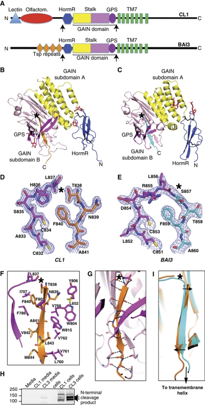
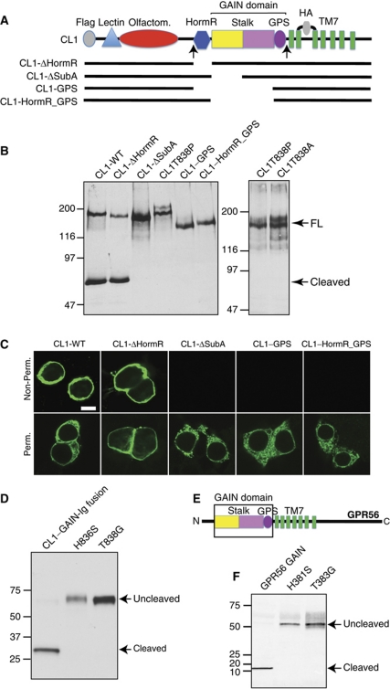
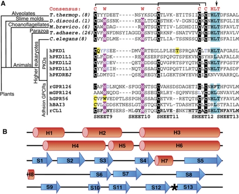
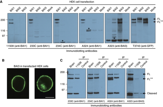
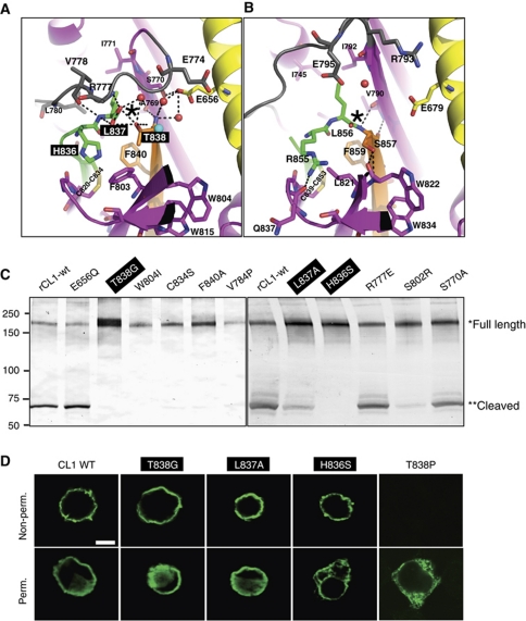
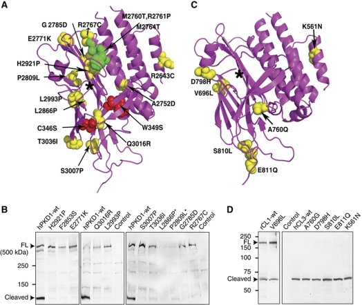
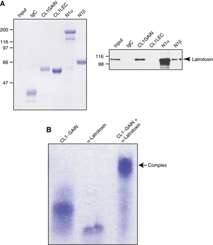
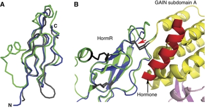
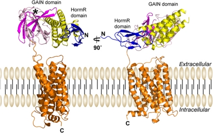
The G protein-coupled receptor (GPCR) Proteolysis Site (GPS) of cell-adhesion GPCRs and polycystic kidney disease (PKD) proteins constitutes a highly conserved autoproteolysis sequence, but its catalytic mechanism remains unknown. Here, we show that unexpectedly the ∼40-residue GPS motif represents an integral part of a much larger ∼320-residue domain that we termed GPCR-Autoproteolysis INducing (GAIN) domain. Crystal structures of GAIN domains from two distantly related cell-adhesion GPCRs revealed a conserved novel fold in which the GPS motif forms five β-strands that are tightly integrated into the overall GAIN domain. The GAIN domain is evolutionarily conserved from tetrahymena to mammals, is the only extracellular domain shared by all human cell-adhesion GPCRs and PKD proteins, and is the locus of multiple human disease mutations. Functionally, the GAIN domain is both necessary and sufficient for autoproteolysis, suggesting an autoproteolytic mechanism whereby the overall GAIN domain fine-tunes the chemical environment in the GPS to catalyse peptide bond hydrolysis. Thus, the GAIN domain embodies a unique, evolutionarily ancient and widespread autoproteolytic fold whose function is likely relevant for GPCR signalling and for multiple human diseases.


Conflict of interest statement

The authors declare that they have no conflict of interest.

Figures

Figure 1
Figure 1
Structures of CL1 and BAI3. (A). Diagram of CL1 and BAI3 showing the domains suggested by the SMART protein domain prediction server. No domain is predicted for the stalk region (coloured yellow and light pink). Note that GPS is defined as a separate domain in the Pfam database (magenta). In contrast, our crystal structures (see B and C) now show that the GPS motif is not an autonomously folded domain, but rather an integral part of the GAIN domain. The stalk region is also part of the GAIN domain. The HormR, TM7, lectin, olfactomedin, and thrombospondin motifs are coloured blue, green, cyan, red, and orange, respectively. The boundaries of the crystal structures are indicated with arrows. (B, C) Cartoon representation of the crystal structure of the HormR and GAIN domains of CL1 (PDB accession code 4DLQ) (B) and BAI3 (PDB accession code 4DLO) (C). The HormR domain is coloured blue, the GAIN subdomain A yellow, the GAIN subdomain B light pink, and the GPS motif, which is part of subdomain B, magenta. The cleaved β-strand of CL1 and the corresponding uncleaved β-strand of BAI3 are coloured orange and cyan, respectively. Disulphide bonds and carbohydrates are shown as black and red sticks, respectively. (D) 2mFoDFc electron density map of the indicated residues around the cleavage site in CL1, contoured at 2σ. (E) 2mFoDFc electron density map of the corresponding uncleaved site in BAI3, also contoured at 2σ. (F) Showing the hydrophobic packing interactions between the cleaved β-strand and the surrounding residues in CL1. (G) Showing the hydrogen bonds between the cleaved β-strand and the surrounding residues in CL1. In all panels, the cleavage site in CL1 and the corresponding peptide bond in BAI3 are marked with a black star. (H) The majority of the extracellular regions of CL1 and CL3 remain attached to the cells upon cleavage. HEK293 cells were transfected with full-length CL1 and CL3 constructs. Equal ratio of cell lysates and media were loaded on the gel and immunoblotted for the N-terminal Flag epitope. (I) The cleaved β-strand of the GAIN domain undergoes a conformational change upon autoproteolysis (the direction of the movement is indicated by arrows).
Figure 2
Figure 2
The GAIN domain is an autoproteolytic domain. (A) Diagram showing the CL1 deletion mutants that we tested in full-length CL1. The constructs were tagged with Flag and Haemagglutinin A (HA) epitopes (grey), both facing the extracellular side. (B) Effect of domain deletions on cleavage of CL1 constructs in HEK293 cells. For the cleavage assay, cell extracts were immunoblotted for the HA epitope. If cleavage at the GPS site occurs, a C-terminal 70 kDa fragment is produced. FL represents full length. (C) Effect of the domain deletions on cell-surface transport of CL1 in HEK293 cells. Transfected HEK293 cells expressing the indicated CL1 proteins were analysed by immunocytochemistry using antibodies to the HA tag; cells were either analysed with or without permeabilization. Cells were visualized by confocal microscopy using an Alexa-488 secondary antibody. (D) A construct encoding only the GAIN domain of CL1 as an Ig-fusion protein is autoproteolysed in transfected HEK293 cells whereas the two autoproteolysis site mutants are not cleaved (blotted for Ig). (E) Schematic domain diagram of GPR56. (F) A construct encoding only the GAIN domain of GPR56 as a myc tagged PDGF receptor transmembrane domain-fusion protein is autoproteolysed whereas the two autoproteolysis site mutants are not cleaved (blotted for myc-epitope).
Figure 3
Figure 3
The GAIN domain is an ancient domain that exists in primitive organisms. The structure of the GAIN domain was used to discover this evolutionary relationship. First, BLAST searches were performed to find other GPS motifs. Then, among this group of proteins, the search pattern was extended by including approximately 400 residues upstream of the GPS motif. These sequences were aligned by PROMALS and the secondary structure patterns corresponding to each sequence were predicted by PSIPRED. Finally, the predicted secondary structure patterns were checked against the crystal structure of the GAIN domain and only those that had the same pattern were identified as GAIN domains. (A) Showing the sequence alignment of GPS motifs from evolutionary ancient organisms (top, Tetrahymena thermohila (gi89298346), Dictyostelium discoideum (gi66815909), Monosiga brevicollis (gi16752408), Trichoplax adhaerens (gi19600360), Caenorhabditis elegans (gi11553241)), PKD homologues (middle), and cell-adhesion GPCRs (bottom). The evolutionary relationship between the ancient organisms is described as a tree. The minimum number of GAIN domains in each organism's genome is indicated in parenthesis. The conserved cysteines, tryptophans, and cleavage site residues are highlighted black, magenta, and cyan, respectively. Disease mutations (ADPKD, cancer, and BFPP) are highlighted yellow. The cleavage site is indicated with an arrow. Disulphide bonds are shown as black lines. (B) Predicted consensus secondary structure pattern for representative ancient GAIN domains, cell-adhesion GPCRs, and PKD-related proteins shown in Supplementary Tables S2 and S3.
Figure 4
Figure 4
BAI3 GAIN domain is functional but its autoproteolysis is not required for surface transport. (A) Immunoblots of the lysates from HEK293 cells transfected with BAI1, BAI2, or BAI3 constructs tagged with mVenus in their C-terminal tails. Sample amounts were adjusted for immunoblotting to obtain comparable signals for BAIs; therefore, unspecific bands exhibit different intensities (black star). Blots were probed with antibodies raised against C-terminal epitopes of BAI1 and BAI3. Only full-length (FL) BAIs (200 kDa) and N-terminally truncated BAIs of unknown physiological relevance (FL**) (∼170 kDa) were detected, but no products of autoproteolytic cleavage (expected mass: ∼100 kDa) were observed. (B) Images of non-permeabilized HEK293 cells transfected with mVenus-tagged BAI3 cDNA demonstrate cell-surface membrane localization of uncleaved BAI3 (scale bar=10 μm). (C) Immunoblots of mouse brain lysates and of brain immunoprecipitates (IPs) obtained with the BAI1-specific antibody 11509 (control: IP without 11509) with the antibodies used in (A). A C-terminal cleavage product of 72 kDa was detected.
Figure 5
Figure 5
Mutagenesis of the CL1 autoproteolysis site. (A) Close-up view of the cleavage site in CL1 (black star). (B) The corresponding site in uncleaved BAI3 (black star). The colour coding is similar to that in Figure 1, except that the β-strands 12, 13, and the top loop are coloured green, orange, and grey, respectively. Residues involved in autoproteolysis (see mutagenesis experiments) are highlighted black. Water molecules and a sulphate ion are shown as red and cyan spheres, respectively. Hydrogen bonds are shown as black dashes. (C) Results of the cleavage assay of CL1 mutants (same assay as described in Figure 2B). (D) Effect of selected cleavage mutations on cell-surface transport of CL1 in HEK293 cells. Uncleaved, but properly cell-surface localized (i.e., folded), mutants are highlighted black.
Figure 6
Figure 6
The effect of ADPKD and cancer mutations on autoproteolysis. (A) The ADPKD substitution mutations (yellow spheres) and gene conversion mutations (green spheres) in the human PKD1 gene; and BFPP substitution mutations (red spheres) in the human GPR56 gene are superposed on the structure of the CL1 GAIN domain. The cleavage site is indicated with a black star. (B) Effect of ADPKD mutations on the cleavage of the full-length hPKD1 construct that has an N-terminal GFP and a C-terminal Flag tags. HEK293 cells were transfected with the wild-type and mutant hPKD1 constructs and cell extracts were immunoblotted for the Flag epitope. A C-terminal 130 kDa fragment is produced following autoproteolysis in the GAIN domain. The approximate size of hPKD1 is indicated. The mutants that poorly expressed are labelled with black stars. (C) The cancer substitution mutations (yellow spheres) in the human CL1 and CL3 genes marked in the structure of the CL1 GAIN domain. (D) Effect of cancer mutations on the cleavage of the full-length rat CL1 and human CL3 that has tags similar to those shown in Figure 2A. Cell extracts were immunoblotted for the HA epitope. A C-terminal 70 kDa fragment is produced following cleavage. FL represents full length.
Figure 7
Figure 7
The GAIN domain of CL1 binds to α-latrotoxin. (A) Ig-fusion proteins of CL1 comprising the lectin domain (CL1LEC), the GAIN domain (CL1GAIN), or control (IgC) were expressed in HEK293 cells and purified on protein A sepharose beads along with Ig-fusion proteins of Neurexin-1α and Neurexin-1β (N1α and N1β). Recombinant myc–α-latrotoxin produced in bacteria was added. Proteins were visualized by Coomassie-blue staining (left) or immunoblotting using anti-myc antibody (right). (B) Native gel showing the formation of a complex of CL1-GAIN domain and the 20 ankyrin repeats of α-latrotoxin in a calcium-independent manner. Both proteins were expressed in insect cells as His-tagged proteins, secreted to the media, and purified to >99% purity by affinity tag purification followed by size-exclusion chromatography.
Figure 8
Figure 8
The HormR domains of cell-adhesion GPCRs exhibit structural similarity to genuine hormone-binding domains. (A) Superposition of the HormR domains of CL1 (blue), BAI3 (grey), and CRFR (green) (PDB ID 3ehu). The structure of the CL1 HormR domain is superposed on the structure of the CRFR HormR domain (green). (B) Binding of a putative hormone (red α-helix) to the CL1 HormR domain based on hormone (corticotrophin releasing factor) binding to CRFR would produce a clash with the GAIN domain. Thus, a conformational change would be required in order to make the putative hormone binding site accessible to the hormone. The colour coding of CL1 is the same as in Figure 1A. Disulphide bonds are shown as black sticks. The disulphide bond close to the hormone binding site is indicated with a red arrow.
Figure 9
Figure 9
The GAIN and HormR domains are in close proximity to GPCR transmembrane helices. The short linker between the GAIN domain and the first GPCR transmembrane helix brings the GAIN and HormR domains close to the transmembrane domain, restricting possible arrangements between the domains. A model of CL1 is shown with a plausible arrangement of the GAIN and HormR domains. The model represents only one of the possible orientations of the GAIN domain with respect to the transmembrane helices. The seven-pass transmembrane domain (orange) was modelled by homology using the crystal structure of the β2 adrenergic GPCR structure (PDB ID 2RH1) (Cherezov et al, 2007; Rosenbaum et al, 2007), and the GAIN and HormR domains were taken from the crystal structure of CL1. The cleavage site is indicated with a black star.

Comment in

References

    1. Adams PD, Afonine PV, Bunkoczi G, Chen VB, Davis IW, Echols N, Headd JJ, Hung LW, Kapral GJ, Grosse-Kunstleve RW, McCoy AJ, Moriarty NW, Oeffner R, Read RJ, Richardson DC, Richardson JS, Terwilliger TC, Zwart PH (2010) PHENIX: a comprehensive python-based system for macromolecular structure solution. Acta Crystallogr D Biol Crystallogr 66: 213–221 - PMC - PubMed
    1. Arcos-Burgos M, Jain M, Acosta MT, Shively S, Stanescu H, Wallis D, Domene S, Velez JI, Karkera JD, Balog J, Berg K, Kleta R, Gahl WA, Roessler E, Long R, Lie J, Pineda D, Londono AC, Palacio JD, Arbelaez A et al. (2010) A common variant of the latrophilin 3 gene, LPHN3, confers susceptibility to ADHD and predicts effectiveness of stimulant medication. Mol Psychiatry 15: 1053–1066 - PubMed
    1. Blobel CP (2005) ADAMs: key components in EGFR signalling and development. Nat Rev Mol Cell Biol 6: 32–43 - PubMed
    1. Bolliger MF, Martinelli DC, Sudhof TC (2011) The cell-adhesion G protein-coupled receptor BAI3 is a high-affinity receptor for C1q-like proteins. Proc Natl Acad Sci USA 108: 2534–2539 - PMC - PubMed
    1. Chae J, Kim MJ, Goo JH, Collier S, Gubb D, Charlton J, Adler PN, Park WJ (1999) The Drosophila tissue polarity gene starry night encodes a member of the protocadherin family. Development 126: 5421–5429 - PubMed

Publication types

MeSH terms