. 2012 Apr 4;31(7):1727-38.
doi: 10.1038/emboj.2012.23. Epub 2012 Feb 14.

A pRNA-induced structural rearrangement triggers 6S-1 RNA release from RNA polymerase in Bacillus subtilis

Affiliations

A pRNA-induced structural rearrangement triggers 6S-1 RNA release from RNA polymerase in Bacillus subtilis

Benedikt M Beckmann et al. EMBO J. .

Abstract

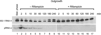
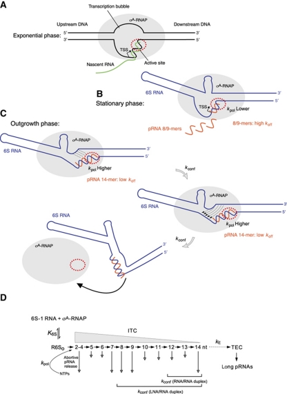
Bacillus subtilis 6S-1 RNA binds to the housekeeping RNA polymerase (σ(A)-RNAP) and directs transcription of short 'product' RNAs (pRNAs). Here, we demonstrate that once newly synthesized pRNAs form a sufficiently stable duplex with 6S-1 RNA, a structural rearrangement is induced in cis, which involves base-pairing between sequences in the 5'-portion of the central bulge and nucleotides that become available as a result of pRNA invasion. The rearrangement decreases 6S-1 RNA affinity for σ(A)-RNAP. Among the pRNA length variants synthesized by σ(A)-RNAP (up to ∼14 nt), only the longer ones, such as 12-14-mers, form a duplex with 6S-1 RNA that is sufficiently long-lived to induce the rearrangement. Yet, an LNA (locked nucleic acid) 8-mer can induce the same rearrangement due to conferring increased duplex stability. We propose that an interplay of rate constants for polymerization (k(pol)), for pRNA:6S-1 RNA hybrid duplex dissociation (k(off)) and for the rearrangement (k(conf)) determines whether pRNAs dissociate or rearrange 6S-1 structure to trigger 6S-1 RNA release from σ(A)-RNAP. A bioinformatic screen suggests that essentially all bacterial 6S RNAs have the potential to undergo a pRNA-induced structural rearrangement.


Conflict of interest statement

The authors declare that they have no conflict of interest.

Figures

Figure 1
Figure 1
B. subtilis 6S-1 RNA forms stable hybrids with pRNA in vitro. (A) In vitro transcription (2 μM B. subtilis RNAP holoenzyme) of pRNA using 3.2 μM 6S-1 RNA as template, either in the presence of all four NTPs (lane 1; each NTP 200 μM) or CTP, GTP and UTP only (lane 2, –ATP). Control lanes: lane 3, as in lane 1 but omission of the 6S-1 RNA template; lanes 4 and 5, chemically synthesized 5′-endlabelled pRNA 8-mer (5′-GUU CGG UC; lane M1) and 14-mer (5′-GUU CGG UCA AAA CU; lane M2) used as size markers. Omission of ATP (lane 2) results in synthesis of short pRNA (8-mers) because 6S-1 RNA encodes four consecutive A residues at pRNA positions 9–12 (see sequence at the bottom); our observation of 9- in addition to 8-mers despite the absence of ATP can be explained by RNAP adding a non-templated residue to the 3′-end. RNA products were separated by 20% denaturing PAGE. (B) Annealing of chemically synthesized pRNAs (8- or 14-mers) to 6S-1 RNA. Asterisks denote the radiolabelled RNA species (6S-1 RNA, 14-mer or 8-mer). Lanes 1 and 2: 5′-endlabelled 6S-1 RNA; in lane 2, the 6S-1 RNA was subjected to the pRNA annealing procedure (for details, see Materials and methods); lane 3: 5′-endlabelled 6S-1 RNA (100 nM unlabelled 6S-1 RNA and trace amounts, <1 nM, of 5′-endlabelled 6S-1 RNA) with pRNA 14-mer (1 μM) pre-annealed as just mentioned; lanes 4–6: annealing of 100 nM unlabelled 6S-1 RNA, trace amounts (<1 nM) of 5′-endlabelled pRNA 14-mer, plus 100 nM (lane 4), 200 nM (lane 5) or 500 nM (lane 6) unlabelled 14-mer; lane 7, as lane 3, but using the pRNA 8-mer instead of the 14-mer; lanes 8–10: as lanes 4–6, but using the pRNA 8-mer. Samples were analysed by 9% native PAGE. (C) In vitro transcription of pRNA from 6S-1 RNA (1 μM) as template using 1 μM RNAP holoenzyme. Lanes 1–3: as lanes 5, 4 and 1, respectively, in (A); lane 4: as lane 3, but subjecting 6S-1 RNA to the annealing procedure before transcription; lanes 5–10: 2 μM (lane 5), 4 μM (lane 6), 20 μM (lane 7) pRNA 14-mer or 2 μM (lane 8), 4 μM (lane 9), 20 μM (lane 10) pRNA 8-mer were subjected to the annealing procedure in the presence of 2 μM 6S-1 RNA in a final volume of 5 μl 1 × TE buffer; then, 2 μl of 5 × activity buffer and 0.5 μl RNAP were added and samples were incubated for 30 min at 37°C before addition of nucleotides and transcription for 1 h at 37°C (final volume 10 μl). RNA products were separated by 20% denaturing PAGE.
Figure 2
Figure 2
Role of pRNA length and pRNA:6S-1 RNA duplex stability. (A) Trace amounts (<1 nM) of 5′-endlabelled 6S-1 RNA and 2.5 μM unlabelled 6S-1 RNA were subjected to the annealing procedure in a volume of 4 μl (see Materials and methods); then, 1 μl of a heparin solution (400 ng/ μl) and 2 μl 5 × activity buffer were added and samples were kept at 37°C; then 1.06 μl RNAP holoenzyme (8 μg/μl) were added and samples were incubated for 30 min at 37°C, followed by addition of 2 μl nucleotide solution (all four NTPs: lanes 2–7; only CTP, GTP and UTP: lanes 9–14) or 2 μl of double-distilled water instead (no NTPs; lanes 15–21) (final volume 10 μl; f.c. RNAP: 2 μM; f.c. 6S-1 RNA: 1 μM; f.c. NTPs: 200 μM each); lanes C: samples incubated for 180 min at 37°C in the absence of NTPs. After transcription at 37°C for the time period indicated above each lane, samples were analysed by 7.5% native PAGE (1 × TBE). (B) Trace amounts (<1 nM) of 5′-endlabelled 6S-1 RNA and 1.7 μM unlabelled 6S-1 RNA, either alone (lane 5) or in the presence of 17 μM pRNA 14-mer (lane 6), 13-mer (5′-GUU CGG UCA AAA C, lane 7) or 12-mer (5′-GUU CGG UCA AAA, lane 8) were annealed in 6 μl 1 × TE buffer and then loaded on a 7.5% native PAA gel (1 × TBE); lanes 1–4: as lanes 5–8, but before gel loading 1 μl of a heparin solution (400 ng/μl) and 2 μl 5 × activity buffer were added and samples were kept at 37°C; then 1.06 μl RNAP holoenzyme (8 μg/μl) was added and samples were incubated for 30 min at 37°C followed by gel loading. (C) Test for annealing of the pRNA 8-mer (lane 1) or an isosequential all-LNA 8-mer (lane 2) to 6S-1 RNA; lanes 3 and 4: as lanes 1 and 2, but after the annealing procedure samples were further incubated with RNAP holoenzyme; for details, see (B) and Materials and methods. Lanes 5–7 illustrate that 6S-1 RNA hybrid complexes with the LNA 8-mer cause the same mobility shift as obtained with the RNA 14-mer.
Figure 3
Figure 3
pRNA induces structural changes in 6S-1 RNA. (A) Structure probing using 5′-endlabelled 6S-1 RNA. Lane 1: 5′-endlabelled 6S-1 RNA directly loaded onto the gel; lane 2: alkaline hydrolysis ladder of 6S-1 RNA; lane 3: limited RNase T1 digest under denaturing conditions; lanes 4 and 5: free 6S-1 RNA subjected to the annealing procedure (lane 5) or not (lane 4) before lead probing; lane 6: 10 pmol 5′-endlabelled 6S-1 RNA and 100 pmol pRNA 14-mer were subjected to the annealing procedure in 6 μl 1 × TE buffer before lead probing. (B) Structure probing using 3′-endlabelled 6S-1 RNA. Lanes 1–3, 6 and 7: as lanes 1–3, 5 and 6, respectively, in (A); lanes 4 and 5: as lanes 6 and 7, but RNase T1 cleavage (under native conditions). Note that lane 1 was taken from a separate gel using the same batch of 3′-endlabelled 6S-1 RNA as in lanes 2–7. (C) Structure probing using 3′-endlabelled 6S-1 RNA. For lanes 1–5, see (B); lanes 6 and 7, cleavage by RNase V1. (D) Structure probing of the 5′-endlabelled circularly permuted 6S-1 RNA (6S-1 cp) variant (see Supplementary Figure S1). Lanes 1–7 correspond to lanes 1–7 in (C); lanes 8 and 9 correspond to lanes 6 and 7 in (B). For details of probing reactions, see Supplementary data. Asterisks in (BD) mark the regions of band compression. (E) Model of the pRNA-induced structural rearrangement of the 6S-1 RNA core region (grey shaded in the 6S-1 RNA secondary structure at the top). The structural rearrangement can be induced by the RNA 14-mer (orange) but also by an all-LNA 8-mer (red; see Supplementary Figure S2) mimicking the 5′-terminal 8 nt of the 14-mer. The orientation of helical elements after the rearrangement is arbitrary, their exact orientation is unknown. RNAfold analysis of the 6S-1 RNA core structure (nt 15–81–(N)4–126–174) predicts that the central bulge collapse occurs when the first nine pRNA-encoding nucleotides are blocked (by constraint folding) for base-pairing with other 6S-1 RNA nucleotides. The three A–U base pairs (U51–53/A153–155) are predicted by RNAfold and mfold in dot plots of suboptimal structures with free energies close to that of the minimum free energy (MFE) structure.
Figure 4
Figure 4
Functional analysis of the U136/U145/A146 mutant 6S-1 RNA unable to form a hairpin in the 3′-portion of the central bulge. (A) 5′-Endlabelled 6S-1 RNA was incubated with σA-RNAP in the presence of NTPs for different time periods as in Figure 2A. Samples were then either loaded on a 7.5% native PAA gel using 1 × TBE as electrophoresis buffer (left panel) or 0.5 × TBE, 160 mM KOAc, 5 mM Mg(OAc)2, 1 mM DTT, pH 8.6 (right panel) as electrophoresis buffer. Lanes C1: samples incubated for 180 min at 37°C in the absence of NTPs; lanes C2: as lanes C1 but without RNAP. For further details, see legend to Figure 2. (B) In vitro transcription was conducted as in Figure 1A, comparing wt (wt 6S-1) and U136/U145/A146 mutant 6S-1 RNA (mut 6S-1) as template; C/G/UTP, NTP mixture lacking ATP. For further details, see legend to Figure 1A. (C) Binding of 5′-endlabelled wt 6S-1 RNA (open squares) and U136/U145/A146 mutant 6S-1 RNA (filled squares) to the σA-RNAP holoenzyme, based on experiments as shown in (A) (right), but in the absence of NTPs (see also Beckmann et al, 2011). Apparent Kd values, based on two independent experiments, were 0.24±0.06 μM for wt 6S-1 RNA and 0.56±0.17 μM for the mutant 6S RNA, as derived from non-linear regression analysis using the equation for a single ligand binding site. Errors, standard errors of the mean (s.e.m.); some error bars are small such that they are masked by the symbol. Similar binding curves were obtained by 7.5% native PAGE in 1 × TBE buffer. The experimental end point for complex formation is lower for the mutant versus wt RNA, suggesting that the fraction of 6S-1 mutant conformers that are unable to bind to RNAP is increased relative to wt 6S-1 RNA. (D) Model of the mutant 6S-1 RNA structure before and after the pRNA-induced structural rearrangement. The three base exchanges are highlighted.
Figure 5
Figure 5
Effect of rifampicin on pRNA synthesis and 6S-1 RNA stability. Analysis of 6S-1 RNA steady-state levels following outgrowth from stationary phase by northern blots of cellular RNA extracted at different time points after induction of outgrowth, either in the absence of rifampicin (lanes 2–9) or in the presence of rifampicin (100 μg/ml; lanes 10–17). Lane 1: RNA extracted from stationary cells immediately before 1:5 dilution in fresh LB medium. An antisense 6S-1 RNA, internally labelled with digoxigenin-UTP (top panel), or a 5′-digoxigenin-labelled LNA/DNA mixmer complementary to the pRNA 14-mer (bottom panel) were used as northern probes; for details see Beckmann et al (2010).
Figure 6
Figure 6
Model of the pRNA length-controlled structural rearrangement of 6S-1 RNA and its release from σA-RNAP. (A) In exponential phase, where cellular 6S-1 RNA concentrations are low, σA-RNAP is primarily engaged in transcription at DNA promoters. (B) During late exponential and stationary phase, 6S-1 RNA levels raise and a substantial fraction of σA-RNAP becomes trapped in complexes with 6S-1 RNA. Here, abortive transcription produces primarily short pRNAs of <10 nt due to a lower kpol for nucleotide addition than under outgrowth conditions (at present, we cannot define at which nucleotide position(s) kpol is critically reduced and to which extent). As a consequence, the high rate constant koff for an RNA/RNA duplex of <10 bp causes the vast majority of pRNAs to dissociate, prompting σA-RNAP to reinitiate transcription. (C) During outgrowth, when nutrients including NTPs are resupplied, the fraction of longer pRNAs (⩾14 nt) increases (Beckmann et al, 2011), because kpol for nucleotide addition increases. When the pRNA transcript reaches a length of 12–14 nt, the hybrid duplex is stable enough to trigger the structural rearrangement (described by the rate constant kconf) that results in dissociation of 6S-1: pRNA hybrids from the enzyme (bottom panel). In the top and middle sketches, the hairpin in the 3′-portion of the central bulge is shown with a curved stem to indicate that it is assumed to be in equilibrium with an open structure (see Figure 3). An inflection of 6S-1 RNA is shown because formation of the hairpin constricts this side of the central bulge. In the top panel, stippled lines depict the progressive disruption of the closing stem, and thin arrows in the middle panel indicate the central bulge collapse. The bottom sketch illustrates the substantial extent of the final structural rearrangement, although the exact relative orientation of the helical elements is unknown. (D) Kinetic scheme adapted from abortive transcription initiation at DNA promoters (Hsu, 2009). Since 6S RNA is thought to mimic an open RNA polymerase–promoter complex (RPo), the equilibrium constant K6S defines formation of the enzyme complex with 6S RNA R6So for simplification, the competition between 6S RNA and DNA promoters as well as other 6S RNAs (such as 6S-2 RNA in B. subtilis) for binding to σA-RNAP is not considered here. Upon addition of NTP substrates, R6So complexes act as initial transcribing complexes (ITCs) releasing abortive transcripts of various length (here 2–14 nt) to different extents (indicated by the length of the vertical arrow). Only pRNAs of ⩾12 nt or isosequential pLNAs ⩾8 nt are capable of inducing the structural rearrangement described by the rate constant kconf. The rate constant for the transition from an ITC to a productive elongation complex (TEC), kE, is thought to be very low if occurring at all with 6S RNAs as transcription templates.

References

    1. Ando Y, Asari S, Suzuma S, Yamane K, Nakamura K (2002) Expression of a small RNA, BS203 RNA, from the yocI-yocJ intergenic region of Bacillus subtilis genome. FEMS Microbiol Lett 207: 29–33 - PubMed
    1. Barrick JE, Sudarsan N, Weinberg Z, Ruzzo WL, Breaker RR (2005) 6S RNA is a widespread regulator of eubacterial RNA polymerase that resembles an open promoter. RNA 11: 774–784 - PMC - PubMed
    1. Beckmann BM, Burenina OY, Hoch PG, Sharma CM, Kubareva EA, Hartmann RK (2011) In vivo and in vitro analysis of 6S RNA-templated short transcripts in Bacillus subtilis. RNA Biol 8: 839–849 - PubMed
    1. Beckmann BM, Grünweller A, Weber MHW, Hartmann RK (2010) Northern blot detection of endogenous small RNAs (∼14 nt) in bacterial total RNA extracts. Nucleic Acids Res 38: e147. - PMC - PubMed
    1. Brownlee GG (1971) Sequence of 6S RNA of E. coli. Nat New Biol 229: 147–149 - PubMed

Publication types

MeSH terms

LinkOut - more resources