

A new whole genome amplification method for studying clonal evolution patterns in malignant colorectal polyps
- PMID: 22334367
- PMCID: PMC3535186
- DOI: 10.1002/gcc.21937
A new whole genome amplification method for studying clonal evolution patterns in malignant colorectal polyps
Abstract
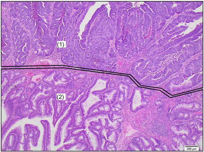
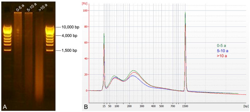
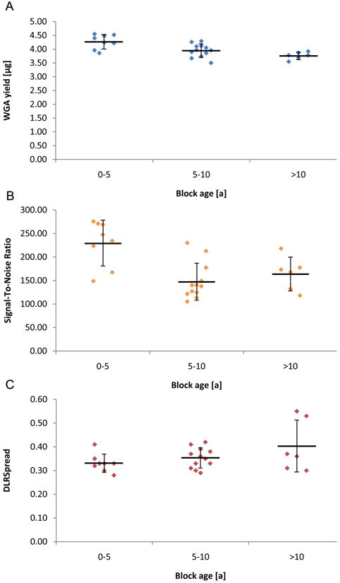
To identify the genetic drivers of colorectal tumorigenesis, we applied array comparative genomic hybridization (aCGH) to 13 formalin-fixed paraffin-embedded (FFPE) samples of early, localized human colon adenocarcinomas arising in high-grade adenomas (so-called "malignant polyps"). These lesions are small and hence the amount of DNA is limited. Additionally, the quality of DNA is compromised due to the fragmentation as a consequence of formalin fixation. To overcome these problems, we optimized a newly developed isothermal whole genome amplification system (NuGEN Ovation® WGA FFPE System). Starting with 100 ng of FFPE DNA, the amplification system produced 4.01 ± 0.29 μg (mean ± standard deviation) of DNA. The excellent quality of amplified DNA was further indicated by a high signal-to-noise ratio and a low derivative log(2) ratio spread. Both, the amount of amplified DNA and aCGH performance were independent of the age of the FFPE blocks and the associated degradation of the extracted DNA. We observed losses of chromosome arms 5q and 18q in the adenoma components of the malignant polyp samples, while the embedded early carcinomas revealed losses of 8p, 17p, and 18, and gains of 7, 13, and 20. Aberrations detected in the adenoma components were invariably maintained in the embedded carcinomas. This approach demonstrates that using isothermally whole genome amplified FFPE DNA is technically suitable for aCGH. In addition to demonstrating the clonal origin of the adenoma and carcinoma part within a malignant polyp, the gain of chromosome arm 20q was an indicator for progression from adenoma to carcinoma.
Copyright © 2012 Wiley Periodicals, Inc.
Conflict of interest statement
Figures


References
-
- Alcock HE, Stephenson TJ, Royds JA, Hammond DW. Analysis of colorectal tumor progression by microdissection and comparative genomic hybridization. Genes Chromosom Canc. 2003;37:369–380. - PubMed
-
- Bardi G, Johansson B, Pandis N, Bak-Jensen E, Orndal C, Heim S, Mandahl N, Andren-Sandberg A, Mitelman F. Cytogenetic aberrations in colorectal adenocarcinomas and their correlation with clinicopathologic features. Cancer. 1993;71:306–314. - PubMed
-
- Bardi G, Johansson B, Pandis N, Mandahl N, Bak-Jensen E, Lindstrom C, Tornqvist A, Frederiksen H, Andren-Sandberg A, Mitelman F, Heim S. Cytogenetic analysis of 52 colorectal carcinomas--non-random aberration pattern and correlation with pathologic parameters. Int J Cancer. 1993;55:422–428. - PubMed
-
- Bardi G, Pandis N, Fenger C, Heim S. Trisomy 7 as the sole cytogenetic aberration in the epithelial component of a colonic adenoma. Cancer Genet Cytogenet. 1995;82:82–84. - PubMed
-
- Benjamini Y, Hochberg Y. Controlling the False Discovery Rate: A Practical and Powerful Approach to Multiple Testing. J R Stat Soc Series B Stat Methodol. 1995;57:289–300.
Publication types
MeSH terms
Substances
Grants and funding
LinkOut - more resources
Full Text Sources
Medical
