. 2012 Apr;21(4):539-52.
doi: 10.1002/pro.2039. Epub 2012 Feb 28.

Substrate recognition mechanism of a glycosyltrehalose trehalohydrolase from Sulfolobus solfataricus KM1

Affiliations

Substrate recognition mechanism of a glycosyltrehalose trehalohydrolase from Sulfolobus solfataricus KM1

Nobuo Okazaki et al. Protein Sci. 2012 Apr.

Abstract

Glycosyltrehalose trehalohydrolase (GTHase) is an α-amylase that cleaves the α-1,4 bond adjacent to the α-1,1 bond of maltooligosyltrehalose to release trehalose. To investigate the catalytic and substrate recognition mechanisms of GTHase, two residues, Asp252 (nucleophile) and Glu283 (general acid/base), located at the catalytic site of GTHase were mutated (Asp252→Ser (D252S), Glu (D252E) and Glu283→Gln (E283Q)), and the activity and structure of the enzyme were investigated. The E283Q, D252E, and D252S mutants showed only 0.04, 0.03, and 0.6% of enzymatic activity against the wild-type, respectively. The crystal structure of the E283Q mutant GTHase in complex with the substrate, maltotriosyltrehalose (G3-Tre), was determined to 2.6-Å resolution. The structure with G3-Tre indicated that GTHase has at least five substrate binding subsites and that Glu283 is the catalytic acid, and Asp252 is the nucleophile that attacks the C1 carbon in the glycosidic linkage of G3-Tre. The complex structure also revealed a scheme for substrate recognition by GTHase. Substrate recognition involves two unique interactions: stacking of Tyr325 with the terminal glucose ring of the trehalose moiety and perpendicularly placement of Trp215 to the pyranose rings at the subsites -1 and +1 glucose.


Figures

Scheme 1
Scheme 1
Figure 1
Figure 1
Stereo view of the overall dimer structure of E283Q mutant GTHase. The dimer is crosslinked by an intermolecular disulfide bridge at Cys298. Domains (A−E) are colored by red, green, blue, yellow, and cyan, respectively. Green mesh represents |FoFc| omit map contoured 3.0 σ around G3-Tre. Tyr325 is stacking with the subsite −1 glucose. The figures were prepared by PyMOL (http://www.pymol.org/).
Figure 2
Figure 2
Extra electron densities observed in the mutant GTHases. (a) Wild-type. (b) E283Q mutant GTHase in complex with maltotriosyltrehalose (G3-Tre). (c) E283Q mutant GTHase in complex with maltoheptaose (G7). (d) D252S mutant GTHase cocrystallized with G3-Tre. |FoFc| maps are colored in green mesh and sugar models fitted to the |FoFc| map are colored in black bonds.
Figure 3
Figure 3
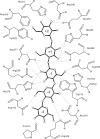
Schematic view of the hydrogen bonding interactions (dotted) between the E283Q mutant GTHase and G3-Tre. Key hydrophobic residues for substrate recognition, Trp215, Phe325, and Phe352 are also included.
Figure 4
Figure 4
Interaction of saccharide in each substrate-binding subsite of GTHase. (a) G3-Tre bound structure of E283Q mutant GTHase (E283Q-G3-Tre). (b) Structure of pig pancreatic α-amylase (PPA) with acarbose. E283Q-G3-Tre structure (thin bonds in green) is superposed as reference model. (c) Structure of E283Q mutant GTHase with G7 (E283Q-G7). (d) Structure of D252S mutant GTHase with G3-Tre (D252S-G3-Tre). (e) The trehalose bound structure of MTHase from Deinococcus radiodurans. E283Q-G3-Tre structure (thin bonds in green) is superposed as reference model.
Figure 5
Figure 5
(a) Pyranose ring distortion observed at the −1 subsite. Thick bonds represent the structure of E283Q mutant MTHase, while thin bonds (colored in yellow) represent the bound G3-Tre. The glucose molecule in the standard conformation is represented by thin cyan bonds. (b) Superposition of D252S with glycerol and E283Q-G3-Tre. D252S, glycerol, and E283Q-G3-Tre are represented in pink, red, and white, respectively. (c) Superposition of D252E and E283Q-G3-Tre. D252E and E283Q-G3-Tre are colored in blue and white, respectively. A rotamer of Glu252 side chain as a model of covalent enzyme-substrate adduct is shown in the thin stick presentation.
Figure 6
Figure 6
(a) ϕ and ψ angle definitions of trehalose and maltose, respectively. (b) Comparison of the +1 and +2 conformation and minimum energy conformations of trehalose and maltose. The lowest (#1) and second lowest (#2) energy conformations of trehalose and maltose are shown. The definition of the ϕ and ψ angles are provided at the bottom left.

Similar articles

Cited by

References

    1. de-Araujo PS. The role of trehalose in cell stress. Braz J Med Biol Res. 1996;29:873–875. - PubMed
    1. Newman YM, Ring SG, Colaco C. The role of trehalose and other carbohydrates in biopreservation. Biotechnol Genet Eng Rev. 1993;11:263–294. - PubMed
    1. Potts M. Desiccation tolerance of prokaryotes. Microbiol Rev. 1994;58:755–805. - PMC - PubMed
    1. Nakada T, Maruta K, Tsusaki K, Kubota M, Chaen H, Sugimoto T, Kurimoto M, Tsujisaka Y. Purification and properties of a novel enzyme, maltooligosyl trehalose synthase, from Arthrobacter sp. Q36. Biosci Biotechnol Biochem. 1995;59:2210–2214. - PubMed
    1. Nakada T, Maruta K, Mitsuzumi H, Kubota M, Chaen H, Sugimoto T, Kurimoto M, Tsujisaka Y. Purification and properties of a novel enzyme, maltooligosyl trehalose trehalohydrolase, from Arthrobacter sp. Q36. Biosci Biotechnol Biochem. 1995;59:2215–2218. - PubMed

Publication types

LinkOut - more resources