. 2012 Feb 28;109(9):3546-51.
doi: 10.1073/pnas.1108718109. Epub 2012 Feb 14.

Protooncogene Ski cooperates with the chromatin-remodeling factor Satb2 in specifying callosal neurons

Affiliations

Protooncogene Ski cooperates with the chromatin-remodeling factor Satb2 in specifying callosal neurons

Constanze Baranek et al. Proc Natl Acad Sci U S A. .

Abstract

First insights into the molecular programs orchestrating the progression from neural stem cells to cortical projection neurons are emerging. Loss of the transcriptional regulator Ski has been linked to the human 1p36 deletion syndrome, which includes central nervous system defects. Here, we report critical roles for Ski in the maintenance of the neural stem cell pool and the specification of callosal neurons. Ski-deficient callosal neurons lose their identity and ectopically express the transcription factor Ctip2. The misspecified callosal neurons largely fail to form the corpus callosum and instead redirect their axons toward subcortical targets. We identify the chromatin-remodeling factor Satb2 as a partner of Ski, and show that both proteins are required for transcriptional repression of Ctip2 in callosal neurons. We propose a model in which Satb2 recruits Ski to the Ctip2 locus, and Ski attracts histone deacetylases, thereby enabling the formation of a functional nucleosome remodeling and deacetylase repressor complex. Our findings establish a central role for Ski-Satb2 interactions in regulating transcriptional mechanisms of callosal neuron specification.


Conflict of interest statement

The authors declare no conflict of interest.

Figures

Fig. 1.
Fig. 1.
Ski is expressed in postmitotic cells of the developing neocortex. (A) Ski immunostaining on coronal brain sections is predominantly detected in the VZ of the dorsal and ventral telencephalon, and in postmitotic cells of the neocortex at E14.5 and E17.5. (B) Higher-magnification images of the rostrodorsal neocortex at E17.5 (boxed region in A Right) show that neurons expressing high levels of Ski are mainly located in the superficial layers of the CP and in layer V as visualized by layer-specific markers Satb2, Ctip2, and Tbr1. (C and D) Ski shows high coexpression with Satb2 in upper layers of the CP and to a minor extent in layer V (yellow in the corresponding overlays) (C), and with Ctip2 in layer V neurons (D). (E) Triple immunostainings for Ski, Satb2, and Ctip2 in higher-magnification images (boxed region in C and D) show that most Ski and Satb2 double-positive cells (yellow in Top, arrow and arrowheads) do not express Ctip2 (blue in Middle, arrowheads). A rare triple-positive cell is depicted (white in Bottom, arrow). (F) Ski and Tbr1 are coexpressed to a minor extent in layer VI neurons. (Scale bars: A, 200 μm; B–F, 20 μm.)
Fig. 2.
Fig. 2.
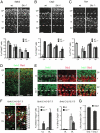
Ski deletion affects Ctip2 and Tbr1 expression patterns in the dorsal telencephalon. (A–C) Satb2 immunostaining of E17.5 coronal brain sections is similar in WT and Ski−/− cortex (A). Ctip2 immunoreactivity has expanded to the superficial layers of the CP in the absence of Ski (B), and fewer cells in the upper layers of the CP express Tbr1 in Ski−/− mice (C). Quantification of Satb2-, Ctip2-, and Tbr1-positive neurons is shown for the superficial layers of the CP, the deep layers V and VI, and the subplate (SP) as a percentage of total DAPI-stained nuclei per field within the respective layer in WT (gray bars) and Ski−/− (black bars). Statistically significant differences were found in the numbers of Ctip2-positive cells (B) and Tbr1-positive cells (C) in the upper layers of the CP. (D and E) Double immunostainings for Satb2 and Ctip2 on E17.5 coronal brain sections in WT and Ski−/− (D). Higher-magnification images reveal ectopic expression of Ctip2 (red) in Satb2-positive cells (green) in Ski mutants (E Lower), whereas Ctip2 expression is absent in Satb2-positive cells of the WT (E Upper). (F) Photomicrographs of neocortical sections show the representative distribution of E14.5 BrdU birth date-labeled, Ctip2-positive cells in WT and Ski−/− (arrows). For the quantification of labeled cells the cortical thickness was divided into 10 equal bins. Bins 1–3 correspond to the upper layers (UL), and bins 7–10 to the deep layers (DL) of the cortical plate. The percentage of BrdU-labeled cells, double positive for Ctip2 in each region (UL, DL) relative to the total number of DAPI-stained nuclei per field, was determined in WT (gray bars) and Ski−/− (black bars; Center and Right). The analysis shows that the numbers of E14.5-born Ctip2-positive cells that populate the UL and DL are significantly increased in the mutant. However, the increase in Ctip2-positive cells in the mutant UL is not due to a precocious generation of these cells, because Ctip2-positive cells born at E12.5 are predominantly found in the DL in both genotypes. (G) Quantitative RT-PCR was performed to determine Ctip2 mRNA levels in WT and Ski−/− cortices at E18.5. Ctip2 values were normalized to HPRT1 mRNA. cDNA from brains of two WT /Ski−/− littermates (experiments 1 and 2) were generated. Results are presented as ratios of Ctip2 levels in Ski−/− and WT, demonstrating ∼1.5- and 1.8-fold induction of Ctip2 in the Ski−/− mutant. (Scale bars: A–D, 50 μm; E and F, 20 μm.) Data are the mean of at least three embryos per genotype. Error bars indicate SD in A–C and SEM in F and G. Student t test, **P ≤ 0.01, ***P ≤ 0.001.
Fig. 3.
Fig. 3.
Ski deletion leads to failure in the formation of the corpus callosum. (A) Immunohistochemistry for the axonal marker L1 on E18.5 coronal brain sections depicts axonal projections forming the corpus callosum. In comparison with WT (Left, arrow), the population of axons crossing the corpus callosum is largely decreased (Center, arrow) or is completely missing (Right, arrow) in Ski−/− embryos. (B) DiI labeling from the neocortex at E18.5 demonstrates that cortical efferent fibers form the corpus callosum in WT but not in Ski−/− embryos (arrows). (C) Double immunohistochemistry for the neuronal marker NeuN and the glial marker GFAP shows the presence of the glial sling (arrow), the glial wedge (asterisk), and the indusium griseum (arrowhead) in WT and Ski−/−. Note that these structures are present in Ski mutants, but the corpus callosum is missing. (Scale bars: 100 μm.)
Fig. 4.
Fig. 4.
Callosal neurons redirect their axons toward subcortical targets in Ski mutants. (A) The placement of DiI crystals in the CP (arrowheads) in WT and Ski−/−. (B) Schematic representation showing the position of DiI crystal placement in the CP. Ncx, neocortex; dTH, dorsal thalamus. (C) DiI placed in the CP retrogradely labels subcerebrally projecting cortical neurons in both WT and Ski−/−. (D) Higher magnifications of WT and Ski−/− cortical plate shown in C. (E and F) Colocalizing the retrogradely labeled neurons with Satb2 shows that the majority of DiI-labeled neurons are Satb2-positive in the Ski−/− mutant (arrowheads in E), whereas more DiI-labeled neurons are Satb2-negative in the WT (arrows in E). The percentage of subcortically projecting neurons that are Satb2-positive is significantly higher in Ski mutants compared with WT (F). (Scale bars: 20 μm.) Error bars indicate SEM. Student t test, **P ≤ 0.01.
Fig. 5.
Fig. 5.
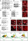
Ski associates with Satb2 and represses Ctip2 transcription in cortical neurons. (A) Lysates from Ski and Satb2-cotransfected (+) and untransfected (–) HEK cells were analyzed by immunoprecipitation (IP) with anti-Ski Ab (IP Ski), anti-Satb2 Ab (IP Satb2), or an unrelated control Ab (IP contr). Western blotting was subsequently performed using Abs against Satb2 (Upper) or Ski (Lower). Note that HEK cells endogenously express low levels of Satb2 (input in Upper), whereas there is no endogenous Ski detectable (input in Lower). (B) Lysates of WT cortical tissue were analyzed by IP with anti-Ski Ab (IP Ski) or an unrelated control Ab (IP contr), followed by immunoblotting using Abs against Satb2. An IP with anti-Ski Ab (IP Ski) with lysates of Ski−/− cortical tissue served as control to demonstrate the specificity of the anti-Ski antibody. Equal input of protein extracts was controlled by Lamin detection. (C) Endogenous Ski–Satb2 complexes were detected in situ in cortical neurons on WT brain sections using PLA. Panels represent magnifications of WT, Ski−/−, and Satb2−/− superficial layers of the CP. The Duolink fluorescent probe 563 (Materials and Methods) was used as a hybridization probe (green), and the nuclei were stained with TO-PRO-3 (red). The Ski–Satb2 complex formation in the WT (Top, arrow) was specific, because there was no signal detectable in the Ski−/− and Satb2−/− (Middle and Bottom). (D and E) Semiquantitative ChIP assay was performed to detect protein occupancy at the Ctip2 locus using WT, Ski−/−, and Satb2−/− cortical tissue from E18.5/P0 pups. A 245-bp fragment was amplified from a previously described Ctip2 regulatory DNA sequence, the matrix attachment region 4 (8) (Fig. S6 A and C). The samples were immunoprecipitated with anti-Ski and anti-Satb2 antibodies (D) or anti-MTA2 and anti-HDAC1 (E). (F and G) Lysates of WT and Ski−/− cortical tissue were analyzed by IP with anti-HDAC Ab (IP HDAC) (F) or anti-MTA2 Ab (IP MTA2) (G) followed by immunoblotting using Abs against Satb2 (F and G). An unrelated Ab was used as control (IP contr) (F and G). Equal input of protein extracts was controlled by Lamin detection (F and G). Note that Satb2–HDAC complex formation is drastically reduced in Ski−/− (F), whereas Satb2–MTA2 complex formation is unaffected in the absence of Ski (G). (H) Using PLA, endogenous protein complex formation was detected in UL neurons in situ as indicated on WT, Ski−/−, and Satb2−/− cortical brain sections (arrows). Note that Satb2–HDAC1 complex formation is absent in UL neurons on Ski−/− sections. (I) Model for Ski function at the Ctip2 locus in callosal projection neurons. Ski is required to assemble a functional NuRD repressor complex containing Satb2, MTA2, and HDAC1 at MAR sites in the Ctip2 locus. In the absence of Ski, Satb2 still binds the regulatory DNA sequences together with MTA2, but recruitment of HDAC1 is impaired. In the absence of Satb2, the NuRD complex is not assembled. Thus, Satb2 and Ski play specific roles in the formation of a functional NuRD complex, and individual loss of these factors prevents transcriptional repression of Ctip2 in callosal projection neurons. (Scale bars: C and H, 5 μm.)

References

    1. Leone DP, Srinivasan K, Chen B, Alcamo E, McConnell SK. The determination of projection neuron identity in the developing cerebral cortex. Curr Opin Neurobiol. 2008;18:28–35. - PMC - PubMed
    1. Molyneaux BJ, Arlotta P, Menezes JR, Macklis JD. Neuronal subtype specification in the cerebral cortex. Nat Rev Neurosci. 2007;8:427–437. - PubMed
    1. Bonnon C, Atanasoski S. c-Ski in health and disease. Cell Tissue Res. 2012;347:51–64. - PubMed
    1. Lyons GE, et al. Protooncogene c-Ski is expressed in both proliferating and postmitotic neuronal populations. Dev Dyn. 1994;201:354–365. - PubMed
    1. Berk M, Desai SY, Heyman HC, Colmenares C. Mice lacking the ski proto-oncogene have defects in neurulation, craniofacial, patterning, and skeletal muscle development. Genes Dev. 1997;11:2029–2039. - PMC - PubMed

Publication types

MeSH terms