. 2012 Apr 27;287(18):14912-22.
doi: 10.1074/jbc.M111.322362. Epub 2012 Feb 13.

β-Actin-binding complementarity-determining region 2 of variable heavy chain from monoclonal antibody C7 induces apoptosis in several human tumor cells and is protective against metastatic melanoma

Affiliations

β-Actin-binding complementarity-determining region 2 of variable heavy chain from monoclonal antibody C7 induces apoptosis in several human tumor cells and is protective against metastatic melanoma

Denise C Arruda et al. J Biol Chem. .

Abstract

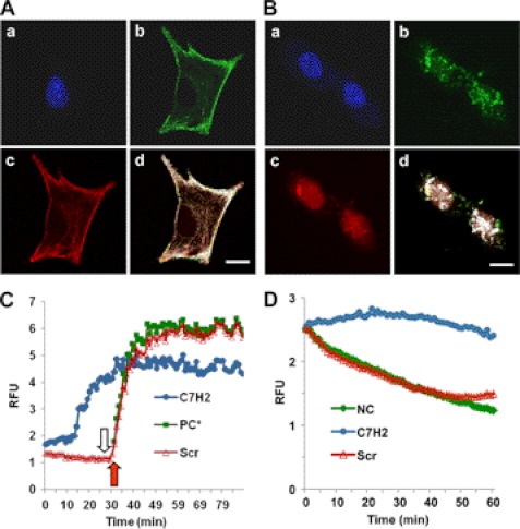
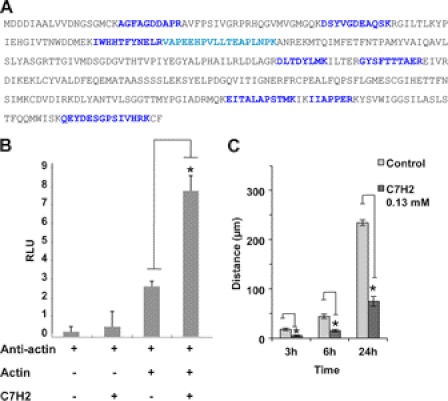
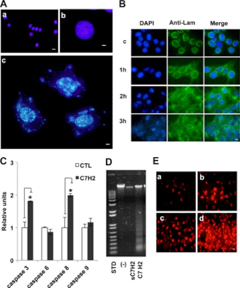
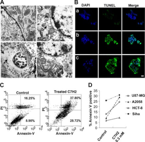
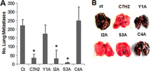
Complementarity-determining regions (CDRs) from monoclonal antibodies tested as synthetic peptides display anti-infective and antitumor activities, independent of the specificity of the native antibody. Previously, we have shown that the synthetic peptide C7H2, based on the heavy chain CDR 2 from monoclonal antibody C7, a mAb directed to a mannoprotein of Candida albicans, significantly reduced B16F10 melanoma growth and lung colony formation by triggering tumor apoptosis. The mechanism, however, by which C7H2 induced apoptosis in tumor cells remained unknown. Here, we demonstrate that C7H2 interacts with components of the tumor cells cytoskeleton, being rapidly internalized after binding to the tumor cell surface. Mass spectrometry analysis and in vitro validation revealed that β-actin is the receptor of C7H2 in the tumor cells. C7H2 induces β-actin polymerization and F-actin stabilization, linked with abundant generation of superoxide anions and apoptosis. Major phenotypes following peptide binding were chromatin condensation, DNA fragmentation, annexin V binding, lamin disruption, caspase 8 and 3 activation, and organelle alterations. Finally, we evaluated the cytotoxic efficacy of C7H2 in a panel of human tumor cell lines. All tumor cell lines studied were equally susceptible to C7H2 in vitro. The C7H2 amide without further derivatization significantly reduced lung metastasis of mice endovenously challenged with B16F10-Nex2 melanoma cells. No significant cytotoxicity was observed toward nontumorigenic cell lines on short incubation in vitro or in naïve mice injected with a high dose of the peptide. We believe that C7H2 is a promising peptide to be developed as an anticancer drug.


Figures

FIGURE 1.
FIGURE 1.
Binding of C7H2 to actin cytoskeleton and actin polymerization. A, co-localization of phalloidin and C7H2 in melanoma cells. B16F10-Nex2 tumor cells were treated with biotinylated C7H2, fixed with paraformaldehyde, and permeabilized with Triton X-100. Cells were stained with DAPI (a), streptavidin-FITC (b), and phalloidin-rhodamine (c) and were examined by confocal microscopy; d, merge, showing co-localization. Scale bar, 20 μm. B, co-localization of DNase I and C7H2 in melanoma cells. B16F10-Nex2 tumor cells were treated with biotinylated C7H2, fixed, and permeabilized as in A. Cells were stained with DAPI (a), streptavidin-FITC (b), and DNase I-Alexa Fluor 594 (c) and were examined by confocal microscopy; d, merge, showing co-localization. Scale bar, 20 μm. C, pyrene G-actin (0.4 mg/ml) added to a 96-well plate. C7H2 at 200 μm (●, blue) and the scramble peptide Scr-C7H2 (△, red) were added, and the fluorescence at 410 nm was measured in kinetic mode. At the end of 30 min (open arrow) or at the same time without previous incubation with peptide (red arrow), the ATP-containing polymerization buffer was added as a positive control (PC*, green). D, pyrene F-actin from stock incubated for 1 h diluted to 0.2 mg/ml and added to a 96-well plate. C7H2 at 200 μm (●, blue) and the scramble peptide Scr-C7H2 (△, red) were added, and the fluorescence at 410 nm in kinetic mode was measured. Negative control, NC (♦, green), incubation with the G-buffer.
FIGURE 2.
FIGURE 2.
Binding of C7H2 to β-actin and inhibition of cell migration. A, B16F10-Nex2 cell lysate was incubated with biotinylated C7H2. Proteins were separated in streptavidin-agarose and eluted with urea. The eluate was separated in SDS-PAGE, and the single band detected was analyzed by mass spectrometry. Peptides from LC MS/MS leading to β-actin identification are shown, over the entire sequence, in blue. B, ELISA plates were coated with BSA with or without peptide C7H2 at 500 μg/ml. β-Actin was added followed by anti-actin antibody and the secondary anti-IgG. A clear binding of β-actin to C7H2 is demonstrated. p < 0.01. C, cells adhering to 12-well plates were treated with 0.13 mm C7H2. After 12 h, wounds across the wells were done, and cell migration was scored at different times. Error bars, S.D.
FIGURE 3.
FIGURE 3.
C7H2 induces nuclear and mitochondrial alterations. A, chromatin condensation seen by Hoechst 33342 staining of A2058 human melanoma cells treated with C7H2 at 0.3 mm for 20 h. Nucleic acid leakage into the cytoplasm is also seen. a (scale bar, 20 μm) and b (scale bar, 80 μm), control cells; c (scale bar, 80 μm), treated cells. Cells were examined by fluorescence microscopy. B, early disintegration of nuclear lamin following 0.3 mm C7H2 treatment of B16F10-Nex2 cells at the indicated times. First column, DAPI nuclear staining; middle column, anti-lamin B1 + B2 antibody, secondary biotinylated anti-mouse IgG and streptavidin-FITC conjugate; third column, merge. Deconvolution microscopy, scale bar, 20 μm. C, B16F10-Nex2 melanoma cells incubated with C7H2 for 40 min induced increased production of caspases 3 and 8 analyzed by colorimetric assay (p < 0.01) but not caspases 6 and 9. D, DNA degradation in A2058 melanoma cells, induced by C7H2 at 0.3 mm for 20 h; a control with scramble peptide is shown and also the standard (STD) DNA ladder pattern fragments. E, abundant anion superoxide produced by B16F10-Nex2 cells treated with 0.3 mm C7H2. a, untreated cells; b, positive control with 5 mm H2O2; c and d, time-dependent anion superoxide production detected with DHE. Scale bar, 20 μm.
FIGURE 4.
FIGURE 4.
TUNEL assay and morphological alterations induced by C7H2. A, transmission electron microscopy of B16F10-Nex2 cells treated with 0.3 mm C7H2 for 2 h. a, control untreated cell; b, peptide-treated cell with detached nuclear membrane, peripheral accumulation of chromatin and altered cytoplasm, swollen and disrupted organelles (arrows); c and d, cells with condensed chromatin and major disintegration of cytoplasmic structures including mitochondria (arrows). B, TUNEL assay in confocal microscopy, showing untreated cells (a), positive control with 1 μg/ml actinomycin-treated cells (b), and 0.3 mm C7H2-treated A2058 melanoma cells for 2 h (c). Scale bar, 20 μm; C, annexin V binding in A2058 melanoma cells treated with 0.3 mm C7H2 for 12 h. propidium iodide labeling is also shown. D, annexin V binding of four different human cancer cells treated with C7H2 for 12 h. Cells were analyzed by FACS.
FIGURE 5.
FIGURE 5.
Antimetastatic effects of peptides. Lung colonization of B16F10-Nex2 cells in a syngeneic system was used to test the protective activity of C7H2 and N-terminal alanine-substituted analogs Y1A, I2A, S3A, or C4A. Protective activity of peptides injected intraperitoneally with doses of 300 μg using the protocol indicated under “Experimental Procedures” is shown. Mice (n = 5/group) were challenged with 5 × 10⁵ syngeneic B16F10-Nex2 melanoma cells (0.1 ml/mouse). *, p < 0.01; error bars, S.D.

References

    1. Bhutia S. K., Maiti T. K. (2008) Targeting tumors with peptides from natural sources. Trends Biotechnol. 26, 210–217 - PubMed
    1. Daffre S., Bulet P., Spisni A., Ehret-Sabatier L., Rodrigues E. G., Travassos L. R. (2008) in Studies in Natural Products Chemistry (Atta-urRahman, ed) pp. 597–691, Elsevier, The Netherlands
    1. Janin Y. L. (2003) Peptides with anticancer use or potential. Amino Acids 25, 1–40 - PubMed
    1. Polonelli L., Magliani W., Conti S., Bracci L., Lozzi L., Neri P., Adriani D., De Bernardis F., Cassone A. (2003) Therapeutic activity of an engineered synthetic killer antiidiotypic antibody fragment against experimental mucosal and systemic candidiasis. Infect. Immun. 71, 6205–6212 - PMC - PubMed
    1. Magliani W., Conti S., Salati A., Arseni S., Ravanetti L., Frazzi R., Polonelli L. (2004) Engineered killer mimotopes: new synthetic peptides for antimicrobial therapy. Curr. Med. Chem. 11, 1793–1800 - PubMed

Publication types

MeSH terms