. 2012 Apr 6;287(15):11891-8.
doi: 10.1074/jbc.M111.338467. Epub 2012 Feb 13.

Phosphorylation of ORC2 protein dissociates origin recognition complex from chromatin and replication origins

Affiliations

Phosphorylation of ORC2 protein dissociates origin recognition complex from chromatin and replication origins

Kyung Yong Lee et al. J Biol Chem. .

Abstract

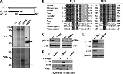
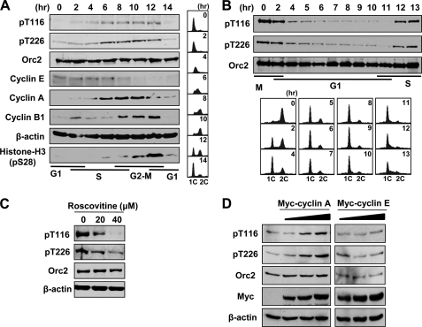
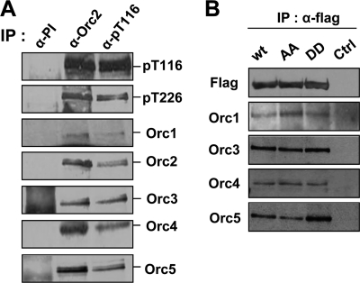
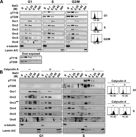
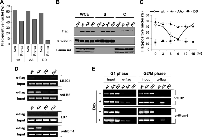
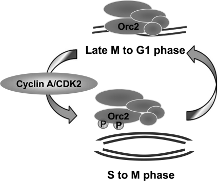
During the late M to the G(1) phase of the cell cycle, the origin recognition complex (ORC) binds to the replication origin, leading to the assembly of the prereplicative complex for subsequent initiation of eukaryotic chromosome replication. We found that the cell cycle-dependent phosphorylation of human ORC2, one of the six subunits of ORC, dissociates ORC2, -3, -4, and -5 (ORC2-5) subunits from chromatin and replication origins. Phosphorylation at Thr-116 and Thr-226 of ORC2 occurs by cyclin-dependent kinase during the S phase and is maintained until the M phase. Phosphorylation of ORC2 at Thr-116 and Thr-226 dissociated the ORC2-5 from chromatin. Consistent with this, the phosphomimetic ORC2 protein exhibited defective binding to replication origins as well as to chromatin, whereas the phosphodefective protein persisted in binding throughout the cell cycle. These results suggest that the phosphorylation of ORC2 dissociates ORC from chromatin and replication origins and inhibits binding of ORC to newly replicated DNA.


Figures

FIGURE 1.
FIGURE 1.
Phosphorylation of human ORC2 at Thr-116 and Thr-226. A, DNA constructs expressing GST-tagged full-length human ORC2 (GST-ORC2), the N-terminal 230 amino acid residues of human ORC2 (GST-ORC2-N), or the C-terminal 347 amino acid residues of human ORC2 (GST-ORC2-C) were transfected into HEK293T cells and incubated in phosphate-free medium containing 200 μCi/ml [32P]orthophosphate for 4 h before harvesting. The 32P and CBB panels represent the autoradiogram and Coomassie Brilliant Blue-stained gel, respectively, of proteins pulled down on glutathione-agarose beads. Ctrl, empty vector; GO2, GST-ORC2; GO2-N, GST-ORC2-N; GO2-C, GST-ORC2-C. B, phosphorylated amino acid residues of human ORC2 determined by mass spectrometry (supplemental Fig. 1) are denoted by asterisks. Alignment of ORC2 protein sequences of human and other eukaryotes was performed using the ClustalW2 program. Ser (S) or Thr (T) in the sequence (S/T)PX(K/R), where X is any amino acid, is potentially phosphorylated by CDK2 (31). C, the indicated proteins, isolated by GST pull-down from HEK293T cells transfected with the corresponding DNA, were analyzed by immunoblot with α-pT116 or α-pT226 antibodies. WT, GST-ORC2; T116A, GST-ORC2 (T116A); T226A, GST-ORC2 (T226A). The lower panels show GST-ORC2 detected by anti-GST-antibody. D, ORC2 protein immunoprecipitated (IP) from cells arrested with double thymidine or nocodazole using a mouse monoclonal anti-ORC2 antibody (α-ORC2; Calbiochem) was immunoblotted using the indicated antibodies. Monoclonal mouse anti-FLAG antibody (α-FLAG; Sigma) was used as control antibody. λ-Phosphatase (λ-PPase) (New England Biolabs) was treated for 30 min at 30 °C. E, HeLa cells were depleted using the following siRNA oligonucleotides (Samchully Pharm): control siRNA (si-GL3), CUUACGCUGAGUACUUCGA; ORC2 siRNA (si-ORC2), GAAGGAGCGAGCGCAGCUU. The depleted cells were immunoblotted using the indicated antibodies.
FIGURE 2.
FIGURE 2.
Cell cycle-dependent phosphorylation of ORC2 by CDK. A, synchronized growth of HeLa cells was achieved by blocking cells in G1/S using double thymidine block. At the indicated times after release into fresh media, cell cycle progression was determined by FACS analysis of propidium iodide-stained cells (right) and by detecting the proteins in immunoblots (left). B, nocodazole (200 ng/ml for 16 h)-treated U2OS cells were collected by mitotic shake-off and released into fresh media. C, HeLa cells were treated with the indicated concentration of roscovitine for 3 h before cell harvest and were analyzed by immunoblot. D, HeLa cells were transfected with 0, 1, 2, and 4 μg of the indicated DNA constructs followed by incubation for 48 h, and the indicated proteins were analyzed by immunoblot.
FIGURE 3.
FIGURE 3.
ORC assembly is not affected by ORC2 phosphorylation. A, HeLa G2/M extracts were prepared by 8-h release from double thymidine block, and immunoprecipitation (IP) was then performed using anti-rabbit preimmune serum (α-PI), anti-ORC2 (α-ORC2), or α-pT116 antibody. B, HEK293 cells were transfected with FLAG-ORC2 (wt), FLAG-ORC2-T116A/226A (AA), or FLAG-ORC2-T116D/226D (DD) expressing DNA empty vector (Ctrl) and were harvested after 48 h. Each lysate was incubated with anti-FLAG antibody for 3 h, and immunoprecipitates were immunoblotted using the corresponding antibody.
FIGURE 4.
FIGURE 4.
Phosphorylation of ORC2 in the S phase dissociates the ORC from chromatin. A, HeLa cells arrested with nocodazole were released for 4 h for G1 phase cells. For S- and G2/M phase cells, the cells arrested with double thymidine block were released for 4 and 8 h, respectively. The FACS profiles of harvested cells are shown in the top panel. The chromatin fractionations of harvested cells were performed as described under “Experimental Procedures.” The indicated proteins were detected by immunoblot analysis. S, soluble fraction; ppt, precipitate after 0.45 m NaCl elution. B, 10 nm calyculin A, as the final concentration, was added to synchronously growing S or G1 phase HeLa cell cultures 30 min prior to cell harvest and during chromatin fractionation (+) or not at all (−). The FACS profiles of each cell sample are shown in the bottom panel.
FIGURE 5.
FIGURE 5.
Phosphorylation of ORC2 dissociates the ORC from chromatin and the replication origin. A, HeLa Tet-On cells expressing FLAG-tagged ORC2 wild type (wt), T116A/T226A (AA) mutant, or T116D/T226D (DD) mutant were incubated in media containing 2 μg/ml doxycycline for 48 h. The cells were fixed with 2% paraformaldehyde and then stained with anti-FLAG antibody, followed by Cy3-conjugated secondary antibody. Pre-extraction was performed using 0.5% Triton X-100 in phosphate-buffered saline for 1 min before fixation (supplemental Fig. 3). The nuclei exhibiting FLAG-positive signal were quantified using ImageJ software and described as the ratio of positive nuclei over total number of nuclei. B, chromatin fractionation was performed as described under “Experimental Procedures.” Proteins were detected by immunoblot analysis. WCE, whole-cell extract; S, soluble fraction; C, chromatin-enriched fraction; Ctrl, HeLa Tet-On cells containing no FLAG-ORC2 gene. C, HeLa Tet-On cells expressing the indicated FLAG-ORC protein, which were arrested at the G1/S boundary using a double thymidine block, were released into fresh media for the indicated time. At 24 h before thymidine release, 2 μg/ml doxycycline was added to the medium. After pre-extraction, cells containing FLAG-positive nuclei were scored and indicated as a ratio over total cells. Three independent experiments were performed. D, asynchronously growing HeLa Tet-On cells expressing the corresponding FLAG-tagged ORC2 were obtained by the addition of 2 μg/ml doxycyline for 48 h before cell harvest. The chromatin-immunoprecipitation using an anti-FLAG antibody was followed by amplification by PCR as described under “Experimental Procedures.” Input DNAs were obtained by PCR of the soluble chromatin fraction. oriLB2, the replication origin in lamin B2; LB2C1, 3.5 kbp upstream from oriLB2; oriMcm4, the replication origin in the Mcm4 upstream promoter region; EX7, exon VII coding region of the Mcm4 gene. E, the G1 or the G2/M phase cells were obtained by 8 h after release of nocodazole or from double-thymidine arrested cells, respectively. FLAG-ORC2 proteins were expressed by the addition of 2 μg/ml doxycyline for 48 h before cell harvest. Then ChIP assays were performed. Dox, doxycycline.
FIGURE 6.
FIGURE 6.
ORC2 phosphorylation regulates the association of the ORC with chromatin and the replication origin.

Similar articles

Cited by

References

    1. Bell S. P., Dutta A. (2002) DNA replication in eukaryotic cells. Annu. Rev. Biochem. 71, 333–374 - PubMed
    1. Diffley J. F. (2004) Regulation of early events in chromosome replication. Curr. Biol. 14, R778–R786 - PubMed
    1. Sclafani R. A., Holzen T. M. (2007) Annu. Rev. Genet. 41, 237–280 - PMC - PubMed
    1. Blow J. J., Dutta A. (2005) Preventing re-replication of chromosomal DNA. Nat. Rev. Mol. Cell Biol. 6, 476–486 - PMC - PubMed
    1. DePamphilis M. L., Blow J. J., Ghosh S., Saha T., Noguchi K., Vassilev A. (2006) Regulating the licensing of DNA replication origins in metazoa. Curr. Opin. Cell Biol. 18, 231–239 - PubMed

Publication types