. 2012 Mar 30;287(14):11174-82.
doi: 10.1074/jbc.M111.309070. Epub 2012 Feb 10.

Synaptic released zinc promotes tau hyperphosphorylation by inhibition of protein phosphatase 2A (PP2A)

Affiliations

Synaptic released zinc promotes tau hyperphosphorylation by inhibition of protein phosphatase 2A (PP2A)

Xu-Ying Sun et al. J Biol Chem. .

Abstract

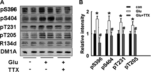
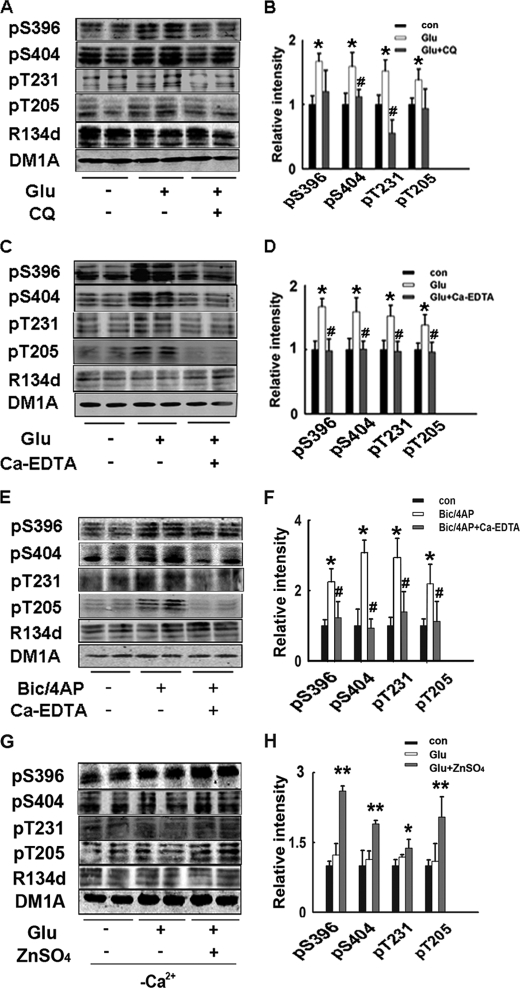
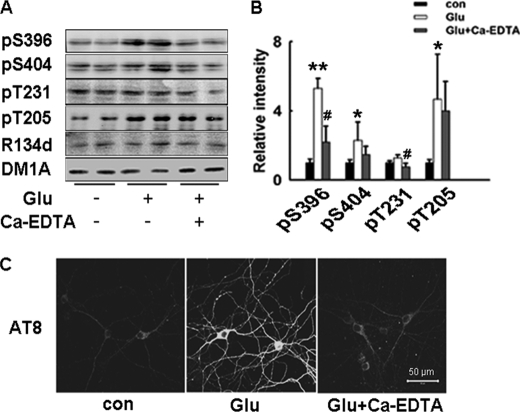
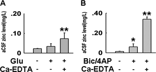
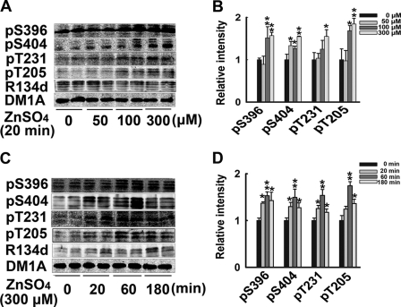
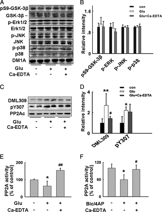
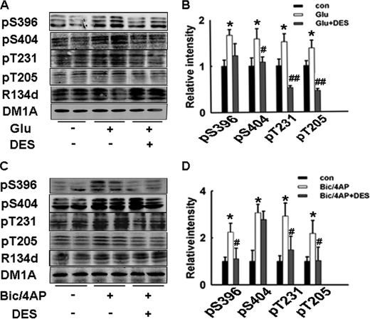
Hyperphosphorylated tau is the major component of neurofibrillary tangles in Alzheimer disease (AD), and the tangle distribution largely overlaps with zinc-containing glutamatergic neurons, suggesting that zinc released in synaptic terminals may play a role in tau phosphorylation. To explore this possibility, we treated cultured hippocampal slices or primary neurons with glutamate or Bic/4-AP to increase the synaptic activity with or without pretreatment of zinc chelators, and then detected the phosphorylation levels of tau. We found that glutamate or Bic/4-AP treatment caused tau hyperphosphorylation at multiple AD-related sites, including Ser-396, Ser-404, Thr-231, and Thr-205, while application of intracellular or extracellular zinc chelators, or blockade of zinc release by extracellular calcium omission almost abolished the synaptic activity-associated tau hyperphosphorylation. The zinc release and translocation of excitatory synapses in the hippocampus were detected, and zinc-induced tau hyperphosphorylation was also observed in cultured brain slices incubated with exogenously supplemented zinc. Tau hyperphosphorylation induced by synaptic activity was strongly associated with inactivation of protein phosphatase 2A (PP2A), and this inactivation can be reversed by pretreatment of zinc chelator. Together, these results suggest that synaptically released zinc promotes tau hyperphosphorylation through PP2A inhibition.


Figures

FIGURE 1.
FIGURE 1.
Increased synaptic activity induces tau hyperphosphorylation at multiple AD-related sites in cultured hippocampal slices. A, rat hippocampal brain slices were incubated with glutamate (100 μm, to increase synaptic activity) for 20 min, with or without the pretreatment of TTX (3 μm, synaptic transmission blocker). Tau phosphorylation levels at Ser-396, Ser-404, Thr-205, and Thr-231 were detected by Western blotting. B, quantitative analysis of the blots in A. Tau phosphorylation levels at different sites were normalized by total tau level recognized by R134d.*, p < 0.05 versus control slices; #, p < 0.05 versus glutamate-treated slices.
FIGURE 2.
FIGURE 2.
Synaptic activity-induced tau hyperphosphorylation is dependent on endogenously released zinc. Rat hippocampal brain slices were incubated with glutamate (100 μm) for 20 min, with or without the pretreatment of membrane-permeable zinc chelator CQ (50 μm) (A) or membrane-impermeable zinc chelator Ca-EDTA (1 mm) (C), tau phosphorylation levels were detected by Western blotting. B and D, quantitative analysis of the blots in A and C. Tau phosphorylation levels were normalized by total tau levels recognized by R134d, *, p < 0.05 versus control slices; #, p < 0.05 versus glutamate-treated slices. E, rat hippocampal brain slices were incubated with Bic/4-AP (50 μm/250 μm, another experimental model to increase synaptic activity) for 3 h, with or without the pretreatment of Ca-EDTA (1 mm). Tau phosphorylation levels were detected by Western blotting. F, quantitative analysis of blots in E, *, p < 0.05 control versus slices; #, p < 0.05 versus Bic/4-AP-treated slices. G, rat hippocampal brain slices were incubated with glutamate (100 μm) for 20 min in aCSF without Ca2+, with or without addition of ZnSO4 (100 μm). Tau phosphorylation levels were detected by Western blotting. H, quantitative analysis of blots in G, *, p < 0.05; **, p < 0.01 versus control slices.
FIGURE 3.
FIGURE 3.
Increased synaptic activity induces zinc-dependent tau hyperphosphorylation in cultured hippocampal neurons. A, rat primary hippocampal neurons were incubated with glutamate (20 μm, with 10 μm glycine) for 20 min, with or without the pretreatment of extracellular zinc chelator Ca-EDTA (1 mm). Tau phosphorylation levels were detected by Western blotting. B, quantitative analysis of blots in A, *, p < 0.05; **, p < 0.01 versus untreated neurons, #, p < 0.05 versus glutamate-treated neurons. C, hippocampal neurons were treated as described in A, then cells were fixed and immunostained with AT-8 (phosphorylated tau at Ser202/Thr205). Scale bar: 50 μm.
FIGURE 4.
FIGURE 4.
Increased synaptic activity induces zinc release and translocation in cultured hippocampal slices. Zinc levels in aCSF after the hippocampal slices incubation were detected by atomic absorption spectrophotometry. A, zinc levels in aCSF after glutamate treatment with or without preincubation of Ca-EDTA (1 mm, membrane impermeable zinc chelator, which prevents translocation of the released zinc); B, zinc levels in aCSF after Bic/4-AP treatment with or without preincubation of Ca-EDTA. *, p < 0.05; **, p < 0.01 versus control group.
FIGURE 5.
FIGURE 5.
Exogenous zinc induces tau hyperphosphorylation. A, rat hippocampal brain slices were incubated with 0, 50, 100, or 300 μm ZnSO4 for 20 min, tau phosphorylation levels at different sites were detected by Western blotting. B, quantitative analysis of blots in A, *, p < 0.05; **, p < 0.01 versus untreated control slices. C, rat hippocampal brain slices were incubated with 300 μm ZnSO4 for 0, 20 min, 60 min, and 180 min. Tau phosphorylation levels at different sites were detected by Western blotting. D, quantitative analysis of blots in C, *, p < 0.05; **, p < 0.01 versus untreated control slices.
FIGURE 6.
FIGURE 6.
Synaptically released zinc inhibits PP2A with no effects on GSK-3β, ERK1/2, JNK, and p38. Rat hippocampal brain slices were incubated with glutamate, with or without the pretreatment of Ca-EDTA. A, total protein levels and active/inactive forms of GSK-3β, ERK1/2, JNK, and p38 were detected by Western blotting. B, quantitative analysis of the blots in A. The phosphorylation levels were normalized by corresponding total protein levels. C, two inactive forms of PP2A (Tyr-307 phosphorylated and Leu-309 demethylated PP2A) levels were detected by Western blotting. D, quantitative analysis of blots in C. Phosphorylation and demethylation levels of PP2A were normalized by total PP2A level. E, PP2A activities in glutamate-treated slices with or without preincubation of Ca-EDTA. *, p < 0.05; **, p < 0.01 versus control slices; #, p < 0.05; ##, p < 0.01 versus glutamate-treated slices. F, rat hippocampal brain slices were incubated with Bic/4-AP, with or without the pretreatment of Ca-EDTA. PP2A activities in slices were detected. *, p < 0.05 versus control slices; #, p < 0.05 versus Bic/4-AP-treated slices.
FIGURE 7.
FIGURE 7.
PP2A agonist DES reverses synaptic activity-induced tau hyperphosphorylation. Rat hippocampal brain slices were incubated with glutamate (100 μm) for 20 min (A), or Bic/4-AP (50 μm/250 μm) for 3 h (C), with or without the pretreatment of DES (10 nm, PP2A activator). Tau phosphorylation levels were detected by Western blotting. B and D, quantitative analysis of the blots in A and C. Tau phosphorylation levels were normalized by total tau levels recognized by R134d. *, p < 0.05 versus control slices; #, p < 0.05; ##, p < 0.01 versus glutamate- or Bic/4-AP-treated slices.

Similar articles

Cited by

References

    1. Mount C., Downton C. (2006) Alzheimer disease: progress or profit? Nat. Med. 12, 780–784 - PubMed
    1. Grundke-Iqbal I., Iqbal K., Quinlan M., Tung Y. C., Zaidi M. S., Wisniewski H. M. (1986) Microtubule-associated protein tau. A component of Alzheimer paired helical filaments. J. Biol. Chem. 261, 6084–6089 - PubMed
    1. Wang J. Z., Liu F. (2008) Microtubule-associated protein tau in development, degeneration and protection of neurons. Prog. Neurobiol. 85, 148–175 - PubMed
    1. Bancher C., Brunner C., Lassmann H., Budka H., Jellinger K., Wiche G., Seitelberger F., Grundke-Iqbal I., Iqbal K., Wisniewski H. M. (1989) Accumulation of abnormally phosphorylated tau precedes the formation of neurofibrillary tangles in Alzheimer's disease. Brain Res. 477, 90–99 - PubMed
    1. Köpke E., Tung Y. C., Shaikh S., Alonso A. C., Iqbal K., Grundke-Iqbal I. (1993) Microtubule-associated protein tau. Abnormal phosphorylation of a non-paired helical filament pool in Alzheimer disease. J. Biol. Chem. 268, 24374–24384 - PubMed

Publication types