. 2012 Mar 30;287(14):10714-26.
doi: 10.1074/jbc.M111.299594. Epub 2012 Feb 10.

Kindlin-3 mediates integrin αLβ2 outside-in signaling, and it interacts with scaffold protein receptor for activated-C kinase 1 (RACK1)

Affiliations

Kindlin-3 mediates integrin αLβ2 outside-in signaling, and it interacts with scaffold protein receptor for activated-C kinase 1 (RACK1)

Chen Feng et al. J Biol Chem. .

Abstract

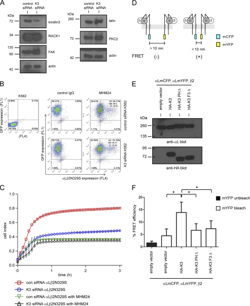
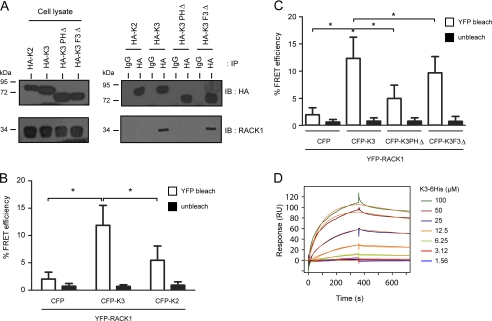
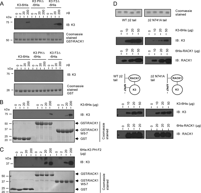
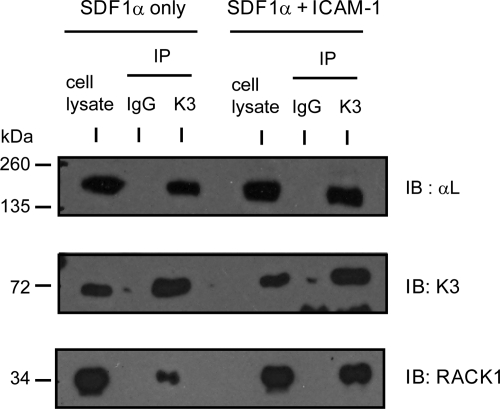
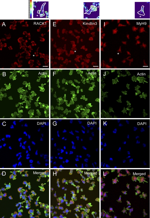
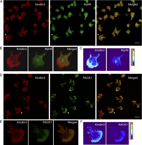
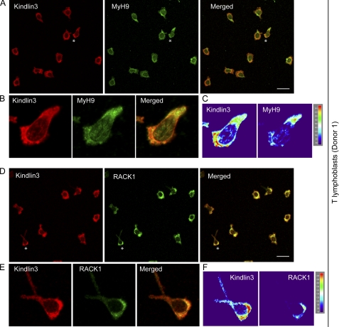
Integrins are heterodimeric type I membrane cell adhesion molecules that are involved in many biological processes. Integrins are bidirectional signal transducers because their cytoplasmic tails are docking sites for cytoskeletal and signaling molecules. Kindlins are cytoplasmic molecules that mediate inside-out signaling and activation of the integrins. The three kindlin paralogs in humans are kindlin-1, -2, and -3. Each of these contains a 4.1-ezrin-radixin-moesin (FERM) domain and a pleckstrin homology domain. Kindlin-3 is expressed in platelets, hematopoietic cells, and endothelial cells. Here we show that kindlin-3 is involved in integrin αLβ2 outside-in signaling. It also promotes micro-clustering of integrin αLβ2. We provide evidence that kindlin-3 interacts with the receptor for activated-C kinase 1 (RACK1), a scaffold protein that folds into a seven-blade propeller. This interaction involves the pleckstrin homology domain of kindlin-3 and blades 5-7 of RACK1. Using the SKW3 human T lymphoma cells, we show that integrin αLβ2 engagement by its ligand ICAM-1 promotes the association of kindlin-3 with RACK1. We also show that kindlin-3 co-localizes with RACK1 in polarized SKW3 cells and human T lymphoblasts. Our findings suggest that kindlin-3 plays an important role in integrin αLβ2 outside-in signaling.


Figures

FIGURE 1.
FIGURE 1.
Kindlin-3 mediates integrin αLβ2 outside-in signaling and promotes integrin αLβ2 micro-clustering. A, cell lysates from K562 stably expressing control siRNA or kindlin-3 (K3)-targeting siRNA were subjected to immunoblotting to assess the expression levels of kindlin-3 and other cytoplasmic proteins. Actin was used as loading control. B, K562 cells were successfully and stably transduced with the lentiviral-based siRNA with GFP as the reporter was transfected with integrin mutant αLβ2N329S. Expression level of integrin αLβ2N329S was determined by staining transfectants with mAb MHM24 followed by APC-conjugated secondary antibody. Two-color flow cytometry analyses were performed. FL1 was for GFP expression, and FL4 was for integrin αLβ2N329S expression. Expression index (EI) was calculated by the percentage of cells gated positive (GP) × geo-mean fluorescence intensity (GM). C, shown is electric cell-substrate impedance sensing measurements of these K562 transfectants on immobilized ICAM-1. The function-blocking mAb MHM24 was included to demonstrate integrin αLβ2-mediated binding specificity. Each data point is the mean ± S.D. (gray bar) of technical triplicates. A representative plot of three independent experiments is shown. Measurements were taken at 1-min intervals, but for clarity cell index is plotted as a function of time at 5-min intervals. D, the drawing illustrates the principle of FRET-based detection of integrin αLβ2 micro-clustering using the αL subunit having a C-terminal fusion of mCFP or mYFP as previously reported (9, 43). E, shown are immunoblots of cell lysates of K562 transfected with integrin subunits αLmCFP, αLmYFP, β2, and HA-tagged kindlin-3 constructs. F, shown are FRET analyses of integrin αLβ2 micro-clustering in K562 transfectants. Each data point represents the mean ± S.D. of ≥40 cells analyzed. *, p < 0.05, Student's t test. A representative plot of three independent experiments is shown.
FIGURE 2.
FIGURE 2.
Kindlin-3 interacts with the scaffold protein RACK1. A, 293T cells transfected with HA-tagged kindlin-2 (K2), HA-tagged kindlin-3 (K3), or its mutants were subjected to immunoprecipitation (IP) analyses. Co-precipitated endogenous RACK1 was detected by immunoblotting (IB). B and C, shown are FRET analyses of COS-7 cells transfected with different CFP constructs and YFP-RACK1. Each data point represents the mean ± S.D. of ≥50 cells analyzed. * p < 0.05, Student's t test. A representative plot of three independent experiments is shown for each panel. D, surface plasmon resonance analyses are shown of K3 and RACK1 interaction. Different concentrations of purified K3-His6 were injected across the surface of GST or GST-RACK-1-coated flow cells in the CM5 sensor chip. The sensorgrams from GST-RACK1 flow cell were double-referenced (41) by subtracting all resultant sensorgrams with the control GST surface (to eliminate nonspecific binding) and blank buffer injections (to eliminate equipment systematic error). The experimental data (colored lines) conformed well to a simple bimolecular model (orange lines) with ka = 0.2 × 10⁴ m−1s−1, kd = 4.1 × 10⁻⁴ s−1, KD = 1.8 μm. Three replicates of different batches of proteins gave an average affinity constant of 1.60 ± 0.08 μm. RU, response units.
FIGURE 3.
FIGURE 3.
Pulldown assays using recombinant kindlin-3 (K3) and RACK1 proteins. A, B, and C, shown are GST pulldown assays using purified recombinant proteins of K3 and RACK1 and their mutants. Immunoblotting (IB) of K3 was performed using commercial anti-kindlin-3 antibody. D, shown are pulldown assays using N-terminal biotin labeled wild-type β2 tail or β2 N741A integrin tail-peptide bound to streptavidin-agarose beads and purified recombinant K3-His6 and His6-RACK1. Coomassie-stained gels of wild-type β2 or β2 N741A integrin tail peptide bound to streptavidin-agarose beads are shown. Top panel, shown are immunoblots of K3-His6 or His6-RACK1 associated with wild-type β2 or β2 N741A integrin tail-peptide. Middle panel, shown is an illustration of ternary complex formation of integrin β2 tail, kindlin-3, and RACK1. Because kindlin-3 interacts with RACK1, a ternary complex of integrin β2 N741A, kindlin-3, and RACK1 could be formed. The dotted line denotes interaction. Bottom panel, shown are immunoblots of K3-His6 associated with wild-type β2 or β2 N741A integrin tail-peptide in the presence of His6-RACK1.
FIGURE 4.
FIGURE 4.
Co-immunoprecipitation of endogenous kindlin-3 with RACK1. SKW3 human T lymphoma cells were seeded into empty or ICAM-1-coated microtiter wells in the presence of SDF-1α. After incubation for 30 min, cells were harvested, and immunoprecipitation (IP) was performed to assess the interaction between kindlin-3 and RACK1. Endogenous kindlin-3 was immunoprecipitated with anti-kindlin-3 mAb (clone 9). An immunoblot (IB) of precipitated kindlin-3 was performed using anti-kindlin-3 (clone 229A). Immunoblots of co-precipitated endogenous RACK1 and integrin αLβ2 were performed using anti-RACK1 mAb (clone B-3) and anti-αL mAb (clone 27), respectively. IgG, control rat IgG.
FIGURE 5.
FIGURE 5.
Immunofluorescence staining of endogenous kindlin-3, RACK1, and MyH9 in SKW3 cells. Cells that adhered to ICAM-1-coated coverslips in the presence of SDF-1α were fixed, permeabilized, and stained for kindlin-3, RACK1, and MyH9 with relevant antibodies as described under “Experimental Procedures.” F-actin and nucleus were stained with Alexa Fluor® 488-conjugated phalloidin and DAPI, respectively. Scale bar, 20 μm. Intensity plots of representative cells are shown (top panel).
FIGURE 6.
FIGURE 6.
Co-localization of endogenous kindlin-3 and RACK1 in SKW3 cells. Shown is immunofluorescence staining of kindlin-3 and MyH9 (A) or kindlin-3 and RACK1 (D) in SKW3 cells that adhered to ICAM-1-coated coverslips in the presence of SDF-1α. B and E, shown are magnified images of selected cells (*). C and F, intensity plots of selected cells (*). Scale bar, 20 μm.
FIGURE 7.
FIGURE 7.
Co-localization of endogenous kindlin-3 and RACK1 in human T lymphoblasts. Shown is immunofluorescence staining of kindlin-3 and MyH9 (A) or kindlin-3 and RACK1 (D) in human T lymphoblasts that adhered to ICAM-1-coated coverslips in the presence of SDF-1α. B and E, shown are magnified images of selected cells (*). C and F, shown are intensity plots of selected cells (*). Scale bar, 20 μm. Cells were from one donor.

Similar articles

Cited by

References

    1. Hynes R. O. (2002) Integrins. Bidirectional, allosteric signaling machines. Cell 110, 673–687 - PubMed
    1. Streuli C. H. (2009) Integrins and cell-fate determination. J. Cell Sci. 122, 171–177 - PMC - PubMed
    1. Shattil S. J., Kim C., Ginsberg M. H. (2010) The final steps of integrin activation. The end game. Nat. Rev. Mol. Cell Biol. 11, 288–300 - PMC - PubMed
    1. Abram C. L., Lowell C. A. (2009) The ins and outs of leukocyte integrin signaling. Annu. Rev. Immunol. 27, 339–362 - PMC - PubMed
    1. Calderwood D. A., Shattil S. J., Ginsberg M. H. (2000) Integrins and actin filaments. Reciprocal regulation of cell adhesion and signaling. J. Biol. Chem. 275, 22607–22610 - PubMed

Publication types

MeSH terms