. 2012 Mar 30;287(14):11037-48.
doi: 10.1074/jbc.M111.310946. Epub 2012 Feb 10.

Down-regulation of B cell receptor signaling by hematopoietic progenitor kinase 1 (HPK1)-mediated phosphorylation and ubiquitination of activated B cell linker protein (BLNK)

Affiliations

Down-regulation of B cell receptor signaling by hematopoietic progenitor kinase 1 (HPK1)-mediated phosphorylation and ubiquitination of activated B cell linker protein (BLNK)

Xiaohong Wang et al. J Biol Chem. .

Abstract

Hematopoietic progenitor kinase 1 (HPK1) is a Ste20-like serine/threonine kinase that suppresses immune responses and autoimmunity. B cell receptor (BCR) signaling activates HPK1 by inducing BLNK/HPK1 interaction. Whether HPK1 can reciprocally regulate BLNK during BCR signaling is unknown. Here, we show that HPK1-deficient B cells display hyper-proliferation and hyper-activation of IκB kinase and MAPKs (ERK, p38, and JNK) upon the ligation of BCR. HPK1 attenuates BCR-induced cell activation via inducing BLNK threonine 152 phosphorylation, which mediates BLNK/14-3-3 binding. Furthermore, threonine 152-phosphorylated BLNK is ubiquitinated at lysine residues 37, 38, and 42, leading to attenuation of MAPK and IκB kinase activation in B cells during BCR signaling. These results reveal a novel negative feedback regulation of BCR signaling by HPK1-mediated phosphorylation, ubiquitination, and subsequent degradation of the activated BLNK.


Figures

FIGURE 1.
FIGURE 1.
HPK1 deficiency enhances BCR-induced cell activation. A, B cell development is not affected in HPK1-deficient mice. Splenocytes from naive mice (n = 3) were stained for different developmental stages of B cells. Transitional 1 (T1, CD23IgDloIgMhiCD21), transitional 2 (T2, CD23+IgDhiIgMhiCD21+), marginal zone (MZ, CD23IgDloIgMhiCD21+), and follicular (FO, CD23+IgDhiIgMloCD21+) B cells were gated from B220+ population from splenocytes. Numbers of B cell subsets were calculated based on total cell numbers of splenocytes and percentages of different B cell subsets. B, in vitro B cell proliferation of WT and HPK1-deficient B cells upon anti-IgM or LPS stimulation. Purified splenic B cells were incubated with anti-IgM or LPS for 40 h. The rate of cell proliferation was determined by the [3H]thymidine incorporation assay. C, HPK1-deficient B cells show increased activation markers upon the stimulation by anti-IgM but not LPS. Purified WT and HPK1-deficient splenic B cells were stimulated with anti-IgM (5–10 μg/ml) or LPS (5 μg/ml) for 6 h (CD69) or 24 h (CD25), respectively. The expression of CD69 (upper panel) and CD25 (lower panel) in these cells was analyzed by flow cytometry.
FIGURE 2.
FIGURE 2.
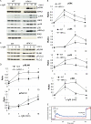
HPK1-deficient B cells show enhanced BCR signaling. Splenic B cells were isolated from WT and HPK1-deficient mice and stimulated with anti-IgM (10 μg/ml) for indicated time periods. The activation of BCR signaling molecules was detected by Western blotting. Western blotting (A) and quantification (B) of phosphorylated and total ERK, p38, JNK, and IKK in WT and HPK1-deficient B cells are shown. Western blotting (C) and quantification (D) of phosphorylated and total BLNK and PLCγ2 in WT and HPK1-deficient B cells are shown. The quantification of immunoblots was performed using Kodak Molecular Imaging Software Version 4.0. The ratios represent the relative phosphorylation levels. Statistical analysis was conducted on the data derived from four independent experiments. *, p < 0.05, Student's t test. E, HPK1-deficient B cells show increased calcium influx upon anti-IgM stimulation. The purified WT and HPK1-deficient splenic B cells were labeled with the calcium indicator Fluo-4. These labeled cells were stimulated with anti-IgM and then with ionomycin. The calcium influx was detected by flow cytometry.
FIGURE 3.
FIGURE 3.
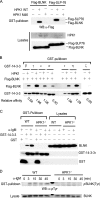
HPK1 induces BLNK/14-3-3 interaction in B cells. A, HPK1 induces BLNK/14-3-3τ interaction in HEK293T cells. FLAG-tagged BLNK was transfected into HEK293T cells with HPK1 WT or kinase-dead mutant (M46). The interaction of FLAG-BLNK/GST-14-3-3 was detected by GST-14-3-3 pulldown assays. FLAG-SLP-76 was included as a positive control for GST-14-3-3 pulldown assays. B, interaction of BLNK with different 14-3-3 isoforms in the presence of HPK1. FLAG-tagged BLNK was transfected into HEK293T cells with HPK1. The interaction of BLNK/14-3-3 isoform was detected by GST pulldown assays. The relative binding affinity of BLNK with 14-3-3 isoforms was calculated by the amount of GST-14-3-3-bound BLNK divided by the amount of GST-14-3-3 input as determined by densitometry analyses. C, BCR stimulation induces BLNK/14-3-3τ interaction in WT but not HPK1-deficient B cells. Splenic B cells were stimulated with anti-IgM for 3 min. Endogenous BLNK/14-3-3τ interaction was pulled down either by GST-14-3-3τ (2nd, 3rd, 5th, and 6th lanes) or by GST alone (1st and 3rd lanes) as controls. D, 14-3-3-associated BLNK is tyrosine-phosphorylated in B cells. WT and HPK1-deficient splenic B cells were stimulated with anti-IgM for indicated time periods and subjected to GST-pulldown assay using GST-14-3-3τ. GST-14-3-3τ-associated BLNK was detected by Western blotting (WB) using anti-phosphotyrosine antibody. Data are representative of three independent experiments.
FIGURE 4.
FIGURE 4.
Thr-152 is phosphorylated by HPK1 and mediates BLNK/14-3-3 interaction. A, schematic diagram of identification of BLNK/14-3-3 binding region. B, interaction of BLNK fragments and 14-3-3 in HEK293T cells was determined by GST-pulldown assays. C, T152A mutation abolishes HPK1-dependent BLNK/14-3-3 interaction in HEK293T cells. Full-length FLAG-BLNK mutants containing S141A, S151A, or T152A mutation were transfected into HEK293T cells with HPK1. The interaction of FLAG-BLNK mutants with GST-14-3-3 was detected by GST-pulldown assays. D, HPK1 induces BLNK Thr-152 phosphorylation in HEK293T cells. FLAG-tagged BLNK WT or T152A mutants were transfected into HEK293T cells with HPK1. FLAG-BLNK was immunoprecipitated with anti-FLAG antibody and Western-blotted with anti-p-BLNK(Thr-152) antibody. E, BCR signaling induces BLNK Thr-152 phosphorylation in WT but not HPK1-deficient B cells. Purified splenic B cells were stimulated with anti-IgM. The phosphorylation of BLNK Thr-152 was detected by Western blotting (WB) with anti-p-BLNK(Thr-152) antibody. Data are representative of three independent experiments.
FIGURE 5.
FIGURE 5.
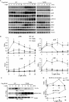
BLNK Thr-152 phosphorylation down-regulates BCR signaling in DT40 B cells. FLAG-tagged BLNK WT, FLAG-tagged BLNK(T152A), and an empty vector control were transfected into BLNK-deficient DT40 mutant chicken B cells. 36 h after transfection, cells were stimulated with anti-chicken IgM (5 μg/ml). Western blotting (A) and quantification (B) of phosphorylated and total ERK, p38, JNK, and IKK in transfected BLNK-deficient DT40 mutant B cells are shown. Statistical analysis was conducted on the data derived from three independent experiments. *, p < 0.05, Student's t test. C, BLNK T152A mutant shows reduced 14-3-3 binding and increased tyrosine phosphorylation in BLNK-deficient DT40 B cells. BLNK-deficient DT40 B cells were transfected as described above. BLNK was immunoprecipitated by anti-FLAG antibody followed by Western blotting (WB) with anti-14-3-3τ or anti-p-BLNK(Y84) antibody. D, quantification of BLNK/14-3-3 interaction and BLNK tyrosine phosphorylation for C.
FIGURE 6.
FIGURE 6.
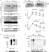
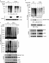
Characterization of BLNK ubiquitination. A, HPK1 deficiency has no discernible effect on the assembly of BLNK- and PLCγ2-interacting complexes. Splenic B cells were isolated from WT and HPK1-deficient mice, stimulated with anti-IgM, and subjected to immunoprecipitation (IP) using anti-BLNK or anti-PLCγ2 antibody. The activation of BLNK and PLCγ2 complexes was determined by Western blotting (WB) with anti-phosphotyrosine antibody. ERK phosphorylation in the whole cell lysate was detected as the control for induction of BCR signaling. B, MG132 stabilizes the Thr-152-phosphorylated BLNK proteins in B cells upon anti-IgM stimulation. WT splenic B cells were cultured in the presence of DMSO or MG132 (20 μm) for 2 h. The cells were then stimulated with anti-IgM. Tyr-84 and Thr-152 phosphorylation of BLNK was determined by Western blot. C, quantification of relative BLNK phosphorylation levels for B. Statistical analysis was conducted on the data derived from three independent experiments. *, p < 0.05, Student's t test. D, detection of BLNK ubiquitination using sequential immunoprecipitation. FLAG-tagged BLNK was transfected into HEK293T cells with HA-ubiquitin. FLAG-BLNK was immunoprecipitated with anti-FLAG antibody (1st IP); half of the 1st anti-FLAG immunoprecipitates was denatured and followed by second round of immunoprecipitation (2nd IP) with anti-FLAG antibody. The ubiquitination of 1st and 2nd anti-FLAG immunoprecipitation was detected by Western blotting analyses with anti-HA antibody. E, BCR stimulation induces endogenous BLNK ubiquitination in WT B cells but not HPK1-deficient B cells. WT and HPK1-deficient B cells were stimulated with anti-IgM stimulation. BLNK was immunoprecipitated by anti-BLNK antibody followed by Western blotting with anti-ubiquitin antibody. Data are representative of three independent experiments.
FIGURE 7.
FIGURE 7.
Thr-152 phosphorylation induces BLNK ubiquitination. A, T152A mutation abolishes HPK1-induced BLNK ubiquitination in HEK293T cells. FLAG-tagged BLNK WT or T152A mutant was transfected into HEK293T cells with HA-ubiquitin and HPK1. BLNK was immunoprecipitated (IP) using anti-FLAG antibody followed by Western blotting (WB) analyses with anti-HA antibody. B, BCR stimulation induces ubiquitination of BLNK WT but not T152A mutant in DT40 B cells. BLNK-deficient DT40 chicken B cells were transfected with FLAG-tagged BLNK WT or T152A mutant with HA-tagged ubiquitin. The transfected cells were either untreated or treated with MG132 for 4 h, followed by stimulation with anti-IgM antibody and immunoprecipitation using anti-FLAG antibody. The ubiquitination of BLNK was detected using anti-ubiquitin (Lys-48) antibody. C, Thr-152-phosphorylated BLNK is ubiquitinated in WT B cells. WT splenic B cells were stimulated with anti-IgM stimulation. BLNK was immunoprecipitated by anti-p-BLNK(Thr-152) followed by Western blotting analyses with anti-ubiquitin or anti-p-BLNK(Thr-152) antibody. HPK1-deficient B cells were included as a negative control. Data are representative of three independent experiments.
FIGURE 8.
FIGURE 8.
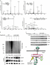
Lys-37, Lys-38, and Lys-42 are BLNK ubiquitination sites in B cells that attenuate BCR signaling. A, MS/MS fragmentation spectra of the tryptic peptides of BLNK contain the ubiquitination modifications of Lys-37, Lys-38, and Lys-42 and phosphorylation of Thr-152. FLAG-BLNK and HA-Ub were transiently transfected into WEHI-231 mouse B cells. The transfected cells were incubated with 25 μm MG132 for 3 h, followed by anti-IgM stimulation for 3 min. FLAG-BLNK were then isolated from the cell lysate using anti-FLAG M2 affinity agarose gel, digested with trypsin, and subjected to LC-MS/MS. B, triple mutations of Lys-37, Lys-38, and Lys-42 residues abolish BLNK ubiquitination in DT40 B cells. BLNK-deficient DT40 B cells were transfected with FLAG-BLNK WT and K37R/K38R/K40R/K42R mutant with HA-ubiquitin, followed by MG132 treatment for 3 h, and then anti-IgM stimulation for 3 min. FLAG-BLNK was immunoprecipitated (IP) using anti-FLAG antibody. Ubiquitination of FLAG-BLNK was detected by Western blotting (WB) analyses using anti-ubiquitin (Lys-48) antibody. C, triple mutations of BLNK Lys-37, Lys-38, and Lys-42 residues enhance ERK, JNK, and IKK activation during BCR signaling. FLAG-BLNK WT and K37R/K38R/K40R/K42R mutant were transfected into BLNK-deficient DT40 B cells, followed by anti-IgM stimulation for indicated time periods. The cell lysates were analyzed by Western blotting for the indicated molecules. GFP was detected as a control for transfection efficiency of BLNK WT and mutant. Data are representative of three independent experiments. D, model of HPK1-mediated negative feedback regulation of BLNK in the attenuation of BCR signaling in B cells. Following BCR stimulation, BLNK is tyrosine-phosphorylated and activated. Activation of BLNK initiates the formation of the BLNK-interacting protein complex, leading to activation of downstream BCR signaling. HPK1 is also activated by interacting with BLNK. The activation of HPK1 in turn induces phosphorylation of BLNK Thr-152, resulting in the binding of 14-3-3. 14-3-3 dimers recruit a putative E3 ubiquitin ligase to BLNK. The putative E3 ligase subsequently induces BLNK ubiquitination at Lys-37, Lys-38, and Lys-42 residues, leading to degradation of the activated BLNK and subsequent attenuation of BCR signaling.

Similar articles

Cited by

References

    1. Yanaba K., Bouaziz J. D., Matsushita T., Magro C. M., St Clair E. W., Tedder T. F. (2008) B-lymphocyte contributions to human autoimmune disease. Immunol. Rev. 223, 284–299 - PubMed
    1. Townsend M. J., Monroe J. G., Chan A. C. (2010) B-cell targeted therapies in human autoimmune diseases: an updated perspective. Immunol. Rev. 237, 264–283 - PubMed
    1. Koretzky G. A., Abtahian F., Silverman M. A. (2006) SLP76 and SLP65. Complex regulation of signaling in lymphocytes and beyond. Nat. Rev. Immunol. 6, 67–78 - PubMed
    1. Datta S. R., Katsov A., Hu L., Petros A., Fesik S. W., Yaffe M. B., Greenberg M. E. (2000) 14-3-3 proteins and survival kinases cooperate to inactivate BAD by BH3 domain phosphorylation. Mol. Cell 6, 41–51 - PubMed
    1. Jang I. K., Zhang J., Gu H. (2009) Grb2, a simple adapter with complex roles in lymphocyte development, function, and signaling. Immunol. Rev. 232, 150–159 - PubMed

Publication types

MeSH terms

Substances