

Mutant protein A30P α-synuclein adopts wild-type fibril structure, despite slower fibrillation kinetics
- PMID: 22334684
- PMCID: PMC3322835
- DOI: 10.1074/jbc.M111.306902
Mutant protein A30P α-synuclein adopts wild-type fibril structure, despite slower fibrillation kinetics
Abstract
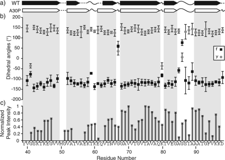
α-Synuclein (AS) is associated with both sporadic and familial forms of Parkinson disease (PD). In sporadic disease, wild-type AS fibrillates and accumulates as Lewy bodies within dopaminergic neurons of the substantia nigra. The accumulation of misfolded AS is associated with the death of these neurons, which underlies many of the clinical features of PD. In addition, a rare missense mutation in AS, A30P, is associated with highly penetrant, autosomal dominant PD, although the pathogenic mechanism is unclear. A30P AS fibrillates more slowly than the wild-type (WT) protein in vitro and has been reported to preferentially adopt a soluble, protofibrillar conformation. This has led to speculation that A30P forms aggregates that are distinct in structure compared with wild-type AS. Here, we perform a detailed comparison of the chemical shifts and secondary structures of these fibrillar species, based upon our recent characterization of full-length WT fibrils. We have assigned A30P AS fibril chemical shifts de novo and used them to determine its secondary structure empirically. Our results illustrate that although A30P forms fibrils more slowly than WT in vitro, the chemical shifts and secondary structure of the resultant fibrils are in high agreement, demonstrating a conserved β-sheet core.
Figures


References
-
- Goedert M. (2001) α-Synuclein and neurodegenerative diseases. Nat. Rev. Neurosci. 2, 492–501 - PubMed
-
- Galvin J. E., Lee V. M., Trojanowski J. Q. (2001) Synucleinopathies: clinical and pathological implications. Arch. Neurol. 58, 186–190 - PubMed
-
- Polymeropoulos M. H., Lavedan C., Leroy E., Ide S. E., Dehejia A., Dutra A., Pike B., Root H., Rubenstein J., Boyer R., Stenroos E. S., Chandrasekharappa S., Athanassiadou A., Papapetropoulos T., Johnson W. G., Lazzarini A. M., Duvoisin R. C., Di Iorio G., Golbe L. I., Nussbaum R. L. (1997) Mutation in the α-synuclein gene identified in families with Parkinson's disease. Science 276, 2045–2047 - PubMed
-
- Krüger R., Kuhn W., Müller T., Woitalla D., Graeber M., Kösel S., Przuntek H., Epplen J. T., Schöls L., Riess O. (1998) Ala30Pro mutation in the gene encoding α-synuclein in Parkinson's disease. Nat. Genet. 18, 106–108 - PubMed
-
- Zarranz J. J., Alegre J., Gómez-Esteban J. C., Lezcano E., Ros R., Ampuero I., Vidal L., Hoenicka J., Rodriguez O., Atarés B., Llorens V., Gomez Tortosa E., del Ser T., Muñoz D. G., de Yebenes J. G. (2004) The new mutation, E46K, of α-synuclein causes Parkinson and Lewy body dementia. Ann. Neurol. 55, 164–173 - PubMed
Publication types
MeSH terms
Substances
Grants and funding
LinkOut - more resources
Full Text Sources
Research Materials
