. 2012 Mar 30;287(14):11018-29.
doi: 10.1074/jbc.M111.318584. Epub 2012 Feb 9.

LytA, major autolysin of Streptococcus pneumoniae, requires access to nascent peptidoglycan

Affiliations

LytA, major autolysin of Streptococcus pneumoniae, requires access to nascent peptidoglycan

Peter Mellroth et al. J Biol Chem. .

Abstract

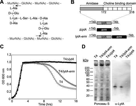
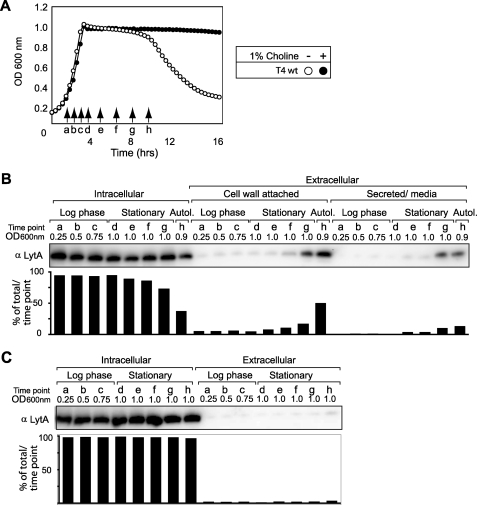
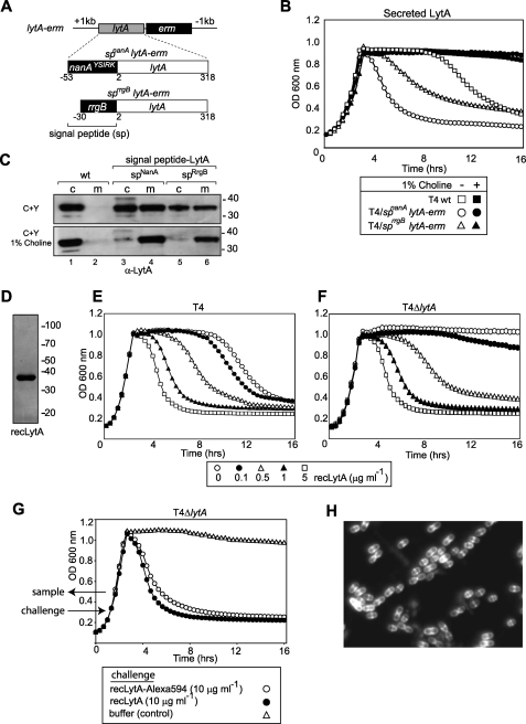
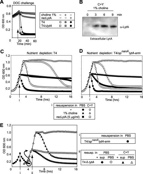
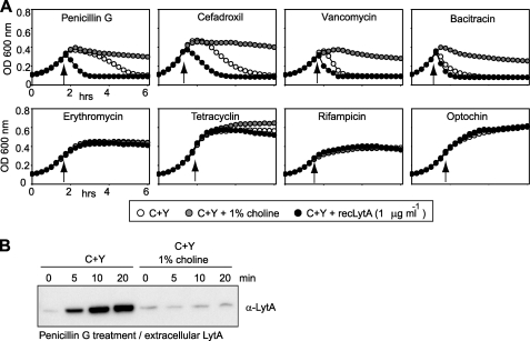
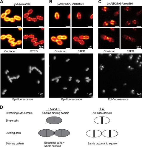
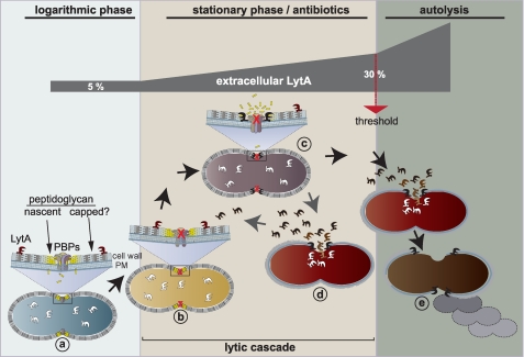
The pneumococcal autolysin LytA is a virulence factor involved in autolysis as well as in fratricidal- and penicillin-induced lysis. In this study, we used biochemical and molecular biological approaches to elucidate which factors control the cytoplasmic translocation and lytic activation of LytA. We show that LytA is mainly localized intracellularly, as only a small fraction was found attached to the extracellular cell wall. By manipulating the extracellular concentration of LytA, we found that the cells were protected from lysis during exponential growth, but not in the stationary phase, and that a defined threshold concentration of extracellular LytA dictates the onset of autolysis. Stalling growth through nutrient depletion, or the specific arrest of cell wall synthesis, sensitized cells for LytA-mediated lysis. Inhibition of cell wall association via the choline binding domain of an exogenously added enzymatically inactive form of LytA revealed a potential substrate for the amidase domain within the cell wall where the formation of nascent peptidoglycan occurs.


Figures

FIGURE 1.
FIGURE 1.
LytA function and genetic strategy. A, LytA is an N-acetylmuramoyl L-alanine amidase that cleaves the peptidoglycan by hydrolyzing the lactyl-amide bond (arrows) that links the peptides and the glycan strands. MurNAc, N-acetyl muramic acid; GlcNAc, N-acetyl glucosamine. B, schematic depicting the LytA amidase and choline binding domains and the genetic organization of the ΔlytA and lytA-erm mutants with respect to the lytA locus. C, growth profiles demonstrating that the TIGR4 (T4) S. pneumoniae mutant T4ΔlytA has a non-autolytic phenotype and that the T4/lytA-erm strain has a slight delay in autolysis compared with T4. D, Ponceau S staining and immunoblot detection of lysates from mid-log cultures (A600 nm = 0.5) of the indicated T4 strains using LytA antisera.
FIGURE 2.
FIGURE 2.
LytA topology distribution in S. pneumoniae. A, growth profile of T4 grown in C+Y media with or without 1% choline chloride. Samples a–h were withdrawn at the indicated time points to investigate the protein topology. B, topology distribution of LytA assayed by immunoblot analysis. Samples a–h (depicted in A) were taken from growing cultures at different OD:s in the log-phase and after 0 h (d), 1.5 h (e), 3 h (f), 4.5 h (g) in stationary phase and at 30 min (h) after autolysis was initiated. The percentage of the total LytA detected intracellularly, cell wall-attached, or secreted into the media was analyzed for each time point. C, topology distribution of LytA from T4 grown in C+Y media with 1% choline assayed by immunoblot analysis. The amount of LytA detected in the intracellular fractions versus extracellular fractions (all present in the media fraction because cell wall attachment was prevented by 1% choline) was analyzed for each time point (a–h).
FIGURE 3.
FIGURE 3.
Extracellular concentration of LytA dictates autolysis onset during stationary phase. A, schematic of the T4 genomic replacements encoding for secreted LytA with a signal peptide from nanA or rrgB expressed from the lytA cognate promoter. B, growth kinetics of the T4 strains secreting LytA in the presence or absence of 1% choline. C, immunoblot analysis of LytA localization in the different strains at an A600 nm = 0.5. Cell-associated LytA (c) was compared with LytA secreted into the media (m) in the absence and presence of 1% choline. D, Coomassie-stained gel of recombinant LytA purified from E. coli. E and F, growth curves displaying how culturing T4 and T4ΔlytA with the indicated amounts of recLytA had a dose-dependent effect on lysis in the stationary phase. G, growth curve of T4ΔlytA cultures challenged with Alexa Fluor 594-labeled recLytA, unlabeled recLytA (10 μg/ml), or buffer at A600 nm = 0.3. A sample was withdrawn at A600 nm = 0.5 after challenge with recLytA-Alexa Fluor 594 for epi-fluorescence live-cell imaging (H).
FIGURE 4.
FIGURE 4.
Disrupting growth during the logarithmic phase may induce sensitivity towards LytA. A, deoxycholate (DOC) challenge during logarithmic growth activates LytA-mediated lysis. Deoxycholate (0.6%) was added at A600 nm = 0.6 to T4 and T4ΔlytA cultures grown in C+Y media in the absence or presence of 1% choline or recLytA (1 μg/ml), and lysis was monitored by a decrease in optical density. B, Deoxycholate perturbs the membrane facilitating the release of intracellular LytA. Shown is an immunoblot analysis displaying the LytA released into the media from T4 cells grown in the presence of 1% choline and sampled at the indicated times after the addition of 0.6% deoxycholate. C and D, nutrient depletion sensitizes pneumococci to LytA-mediated lysis. Cultures of T4 (C) and T4/spnanAlytA-erm (D) were grown in C+Y media (● and ○) or C+Y supplemented with 1% choline (▴ and △) or recLytA (5 μg/ml) (■ and □). At A600 nm ≈ 0.6 (arrows), bacteria was sedimented and resuspended in PBS (●, ▴, and ■) or C+Y (○, △, and □) with or without 1% choline or recLytA. E, LytA activation does not happen at the enzymatic level. T4/spnanAlytA-erm (●) was grown to A600 nm nm ≈ 0.5 (arrow), centrifuged, and resuspended in PBS. When the culture reached A600 nm ≈ 0.1, it was centrifuged, and the filtered supernatant was included in the resuspension mixture of T4ΔlytA cultures that were sedimented at A600 nm ≈ 0.5 and resuspended in 300 μl of PBS or C+Y (plus the 100 μl from the lysed T4/spnanAlytA-erm supernatant).
FIGURE 5.
FIGURE 5.
Cell wall synthesis inhibition during logarithmic growth enables LytA to lyse pneumococci. A, growth profiles of T4R cultures grown in C+Y in the absence or presence of recLytA (1 μg/ml) or 1% choline and challenged with the indicated antibiotics at A600 nm = 0.28 (arrow). B, penicillin does not perturb the bacterial membrane. Shown is the immunoblot analysis of the extracellular amount of LytA following the penicillin G treatment of T4R cultures grown in the absence or presence of 1% choline.
FIGURE 6.
FIGURE 6.
A substrate for LytA adjacent to the equatorial plane. A and B, confocal, STED, and epi-fluorescence images of T4ΔlytA cells from the logarithmic phase stained with Alexa Fluor 594-labeled LytA and LytA[H26A]. C, amidase-bound LytA. Mid-logarithmic phase T4ΔlytA cells were incubated with LytA[H26A]-Alexa Fluor 549 and then consecutively washed and incubated with 1% choline in C+Y to dissociate the choline-bound LytA. D, summary of A–C treatments and LytA staining patterns.
FIGURE 7.
FIGURE 7.
Model for LytA activation. a, a small amount (∼5%) of LytA is cell wall-associated during the logarithmic phase but cannot execute any lytic activity, whereas growth and cell wall synthesis by the plasma membrane (PM)-associated penicillin-binding proteins (PBPs) is in progress. We hypothesize that the inability of LytA to induce lysis is due to capped mature peptidoglycan. b, upon entry into the stationary phase or through challenge with cell wall-targeting antibiotics, the cell wall synthesis machinery becomes inactive, potentially exposing the nascent and yet uncapped peptidoglycan (c), making it available for LytA to initiate cell wall hydrolysis. d, cell wall degradation leads to lysis and release of LytA from the cytoplasmic pool. The newly released LytA can bind to neighboring cells in stages b or c, and the sequential steps b–d constitute a lytic cascade that is initiated at the entry into the stationary phase, causing an accumulation of extracellular LytA. Autolysis is triggered when extracellular LytA reaches ∼30% of total LytA (or ∼0.5 μg/ml), leading to excessive lysis of most cells in the culture (e).

Similar articles

Cited by

References

    1. Neufeld F. (1900) Über eine specifische bakteriolytische Wirkung der Galle. Z. Hyg. Infektionskr. 34, 454–464
    1. Avery O. T., Cullen G. E. (1923) Studies on the Enzymes of Pneumococcus. IV. Bacteriolytic Enzyme. J. Exp. Med. 38, 199–206 - PMC - PubMed
    1. Goebel W. F., Avery O. T. (1929) A Study of Pneumococcus Autolysis. J. Exp. Med. 49, 267–286 - PMC - PubMed
    1. Tomasz A., Albino A., Zanati E. (1970) Multiple antibiotic resistance in a bacterium with suppressed autolytic system. Nature 227, 138–140 - PubMed
    1. Tomasz A., Waks S. (1975) Mechanism of action of penicillin. Triggering of the pneumococcal autolytic enzyme by inhibitors of cell wall synthesis. Proc. Natl. Acad. Sci. U.S.A. 72, 4162–4166 - PMC - PubMed

Publication types

MeSH terms

LinkOut - more resources