. 2012 Mar 23;287(13):10368-10378.
doi: 10.1074/jbc.M111.320697. Epub 2012 Feb 9.

The transmembrane prolines of the mitochondrial ADP/ATP carrier are involved in nucleotide binding and transport and its biogenesis

Affiliations

The transmembrane prolines of the mitochondrial ADP/ATP carrier are involved in nucleotide binding and transport and its biogenesis

Marion Babot et al. J Biol Chem. .

Abstract

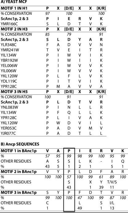
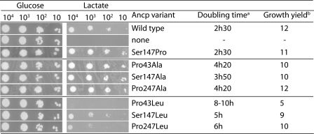
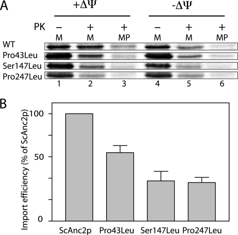
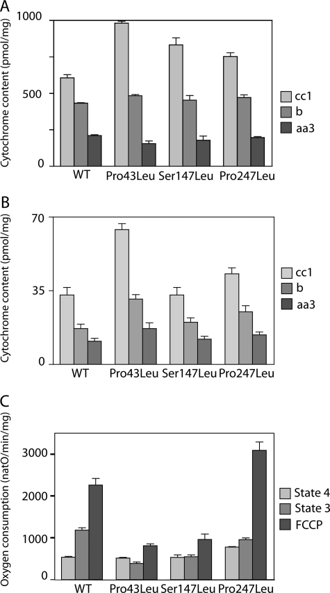
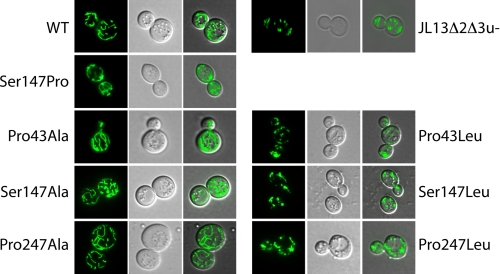
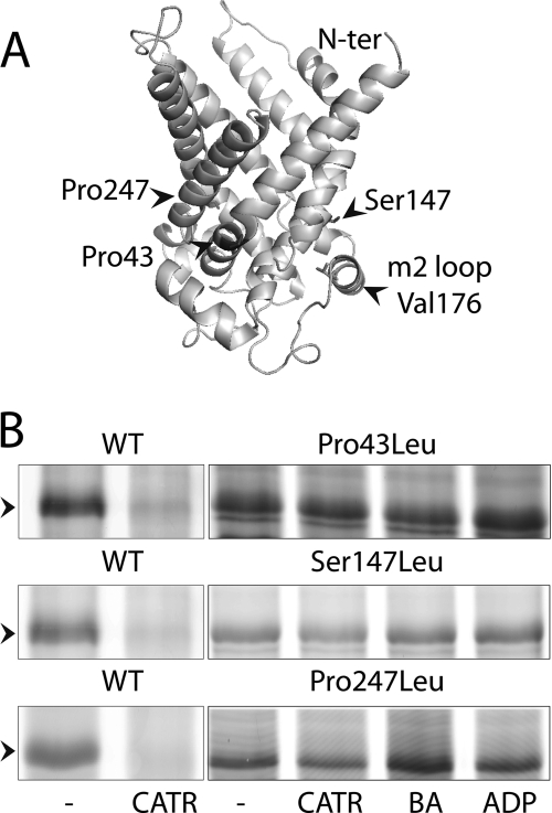
The mitochondrial ADP/ATP carrier (Ancp) is a paradigm of the mitochondrial carrier family, which allows cross-talk between mitochondria, where cell energy is mainly produced, and cytosol, where cell energy is mainly consumed. The members of this family share numerous structural and functional characteristics. Resolution of the atomic structure of the bovine Ancp, in a complex with one of its specific inhibitors, revealed interesting features and suggested the involvement of some particular residues in the movements of the protein to perform translocation of nucleotides from one side of the membrane to the other. They correspond to three prolines located in the odd-numbered transmembrane helices (TMH), Pro-27, Pro-132, and Pro-229. The corresponding residues of the yeast Ancp (Pro-43, Ser-147, and Pro-247) were mutated into alanine or leucine, one at a time and analysis of the various mutants evidenced a crucial role of Pro-43 and Pro-247 during nucleotide transport. Beside, replacement of Ser-147 with proline does not inactivate Ancp and this is discussed in view of the conservation of the three prolines at equivalent positions in the Ancp sequences. These prolines belong to the signature sequences of the mitochondrial carriers and we propose they play a dual role in the mitochondrial ADP/ATP carrier function and biogenesis. Unexpectedly their mutations cause more general effects on mitochondrial biogenesis and morphology, as evidenced by measurements of respiratory rates, cytochrome contents, and also clearly highlighted by fluorescence microscopy.


Figures

FIGURE 1.
FIGURE 1.
Conservation of the mitochondrial carrier signature sequence (A) among the 34 members of the S. cerevisiae MCF and (B) among the mitochondrial ADP/ATP carriers. Sequences were aligned using ClustalX (46). In A, the consensus sequences of the three motives (one-letter amino acid code) are shown, and shown below, the carriers for which the sequences are divergent. ScAnc1p, -2, and -3, mitochondrial ADP/ATP carrier isoforms 1, 2, and 3 (locus tags YR056c, YBL030c, YBR085w). The locus tags are defined in the Saccharomyces Genome Database. B, residues i-1 and i-2 of the consensus prolines are also represented. Occurrences of the residues are indicated as percent of the 114 Ancp sequences analyzed (30). The amino acid immediately after the motif 3 proline corresponds to Trp instead of Phe or Leu in 4% of the Ancp sequences.
FIGURE 2.
FIGURE 2.
Leucine in place of proline is more deleterious to yeast growth than alanine. JL1Δ2Δ3u was transformed with the pRS314 plasmid containing no insert (no Ancp) or the indicated ScAnc2p variant encoding gene. Transformants were cultivated at 28 °C for 2 days on SC-W minimum medium (Glucose) or 4 days on lactate-containing rich medium (Lactate). A600 nm was measured every 4 h during 80–300 h in lactate-rich liquid media to determine doubling time (hours) and growth yield (A600 nm at the stationary phase of the cultures).
FIGURE 3.
FIGURE 3.
In vitro import of the leucine variants into isolated yeast mitochondria. A, [35S]methionine-labeled precursors were incubated with isolated yeast mitochondria, either energized (+ΔΨ) or deenergized (−ΔΨ), for 30 min at 25 °C. Mitochondria (lanes 1, 2, 4, and 6) or mitoplasts (MP, lanes 3 and 6) were then treated with proteinase K (PK, 100 μg/ml; lanes 2, 3, 5, and 6) for 5 min at 4 °C. They were further isolated by centrifugation, washed, and resuspended in sample buffer before SDS-PAGE. Lane 1 corresponds to 10% of the amount of radiolabeled precursor added to each import assay. B, import efficiency for the leucine variants relative to the WT (ScAnc2p in mitoplast after PK digestion/ScAnc2p before the import reaction).
FIGURE 4.
FIGURE 4.
Mitochondria (A) and cell (B) cytochrome contents and mitochondrial respiration rate (C) of the leucine variants. The cytochrome contents are expressed in picomole/mg of proteins (A) or picomole/mg dry weight (B). Oxygen consumption (natO/min/mg) was measured in the presence of NADH (0.2–5 mm) without (state 4) or with 100 μm ADP (state 3). Maximal rate was measured in the presence of 1 μm FCCP.
FIGURE 5.
FIGURE 5.
The mitochondrial morphology is modified in the leucine variant cells. Cells expressing the wild-type or the variants were transformed with a plasmid encoding GFP (S65T) targeted to mitochondria to visualize the mitochondrial network. Cells were cultivated in a selective galactose-containing medium until A600 nm reached a value of 2–4. GFP was visualized with an epifluorescence microscope (Leica Microsystems DM-LB) under an oil immersion objective. WT stands for wild type ScAnc2p.
FIGURE 6.
FIGURE 6.
A, the three-dimensional structure of ScAnc2p was modeled as described in David et al. (12). B, EMA labeling of V176C in the leucine variants. Mitochondria were isolated from ScAnc2pV176C non-mutated (WT) or mutated (P43L, S147L, and P247L). Frozen-thawed mitochondria were incubated (8 mg of proteins/ml) with no ligand (none) or with 20 μm CATR or 20 μm BA or 20 μm ADP for 15 min on ice. EMA labeling (200 μm) was performed for 30 min on ice in the dark. The reaction was stopped with 20 mm DTT. Samples were subjected to SDS-PAGE and fluorescence was visualized at 532 nm. The position of ScAnc2CLpV176C, indicated by an arrow, was controlled by immunoblotting after transfer of the gel onto a nitrocellulose membrane.

Similar articles

Cited by

References

    1. Pebay-Peyroula E., Dahout-Gonzalez C., Kahn R., Trézéguet V., Lauquin G. J.-M., Brandolin G. (2003) Structure of mitochondrial ADP/ATP carrier in complex with carboxyatractyloside. Nature 426, 39–44 - PubMed
    1. Morozzo Della Rocca B., Miniero D. V., Tasco G., Dolce V., Falconi M., Ludovico A., Cappello A. R., Sanchez P., Stipani I., Casadio R., Desideri A., Palmieri F. (2005) Substrate-induced conformational changes of the mitochondrial oxoglutarate carrier. A spectroscopic and molecular modelling study. Mol. Membr. Biol. 22, 443–452 - PubMed
    1. Tonazzi A., Giangregorio N., Palmieri F., Indiveri C. (2005) Relationships of cysteine and lysine residues with the substrate binding site of the mitochondrial ornithine/citrulline carrier. An inhibition kinetic approach combined with the analysis of the homology structural model. Biochim. Biophys. Acta 1718, 53–60 - PubMed
    1. Kunji E. R., Robinson A. J. (2006) The conserved substrate binding site of mitochondrial carriers. Biochim. Biophys. Acta 1757, 1237–1248 - PubMed
    1. Perchiniak E., Lawrence S. A., Kasten S., Woodard B. A., Taylor S. M., Moran R. G. (2007) Probing the mechanism of the hamster mitochondrial folate transporter by mutagenesis and homology modeling. Biochemistry 46, 1557–1567 - PubMed

Publication types

MeSH terms

Substances

LinkOut - more resources