. 2012 Mar 30;287(14):11516-25.
doi: 10.1074/jbc.M111.329219. Epub 2012 Feb 9.

Rapid aquaporin translocation regulates cellular water flow: mechanism of hypotonicity-induced subcellular localization of aquaporin 1 water channel

Affiliations

Rapid aquaporin translocation regulates cellular water flow: mechanism of hypotonicity-induced subcellular localization of aquaporin 1 water channel

Matthew T Conner et al. J Biol Chem. .

Abstract

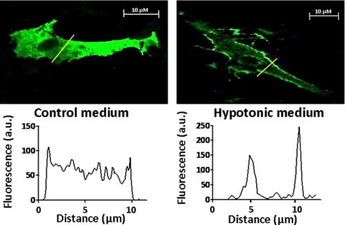
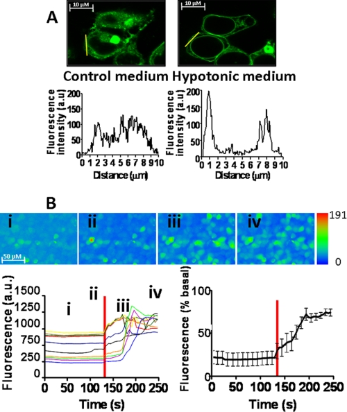
The control of cellular water flow is mediated by the aquaporin (AQP) family of membrane proteins. The structural features of the family and the mechanism of selective water passage through the AQP pore are established, but there remains a gap in our knowledge of how water transport is regulated. Two broad possibilities exist. One is controlling the passage of water through the AQP pore, but this only has been observed as a phenomenon in some plant and microbial AQPs. An alternative is controlling the number of AQPs in the cell membrane. Here, we describe a novel pathway in mammalian cells whereby a hypotonic stimulus directly induces intracellular calcium elevations through transient receptor potential channels, which trigger AQP1 translocation. This translocation, which has a direct role in cell volume regulation, occurs within 30 s and is dependent on calmodulin activation and phosphorylation of AQP1 at two threonine residues by protein kinase C. This direct mechanism provides a rationale for the changes in water transport that are required in response to constantly changing local cellular water availability. Moreover, because calcium is a pluripotent and ubiquitous second messenger in biological systems, the discovery of its role in the regulation of AQP translocation has ramifications for diverse physiological and pathophysiological processes, as well as providing an explanation for the rapid regulation of water flow that is necessary for cell homeostasis.


Figures

FIGURE 1.
FIGURE 1.
AQP1 subcellular localization in fixed, primary rat astrocytes. Immunocytochemistry showing expression profiles of endogenous AQP1 in primary rat astrocytes. Cells shown in each representative image are different fixed cells from the same subculture exposed to different osmotic conditions. Fluorescence amplitudes (a.u., arbitrary units) along the line scans (in yellow on the image) are displayed graphically below each image. RME is calculated from at least five line scan analyses performed in triplicate on a minimum of three independent experiments. Control medium and hypotonic medium are DMEM and F12 at a ratio of 1:1 with osmolalities of 322–374 mosm/kg H2O and 107–125 mosm/kg H2O (diluted with water), respectively. All images and analyses shown are a single representation contributing to the mean value quoted in the text and in Table 1.
FIGURE 2.
FIGURE 2.
Regulation of hypotonicity-induced increase in cell volume by AQP1 translocation in HEK293 cell. Representative images showing x-y surface area (SA) estimation of cell volume. The binary image and surface area are calculated from the z-stack plane at the maximum area. All measurements were taken at 48 h post-transfection. A, HEK293 cells transfected with wild-type AQP0-GFP. B, HEK293 cells transfected with wild-type AQP1-GFP. C, HEK293 cells transfected with the T157A/T239A translocation-deficient mutant of AQP1-GFP. Control medium and hypotonic medium are DMEM with osmolalities of 322–374 mosm/kg H2O and 107–125 mosm/kg H2O (diluted with water), respectively. Distribution profiles along the line scans (in yellow on the image) are displayed graphically by each image indicating AQP distribution in control and hypotonic medium (a.u. is arbitrary units). All images and analyses shown are a single representation contributing to the mean value quoted in the text and in Table 1.
FIGURE 3.
FIGURE 3.
Hypotonicity-induced intracellular Ca2+ elevations and AQP1 translocation in HEK293 cells. A, subcellular localization of AQP1-GFP fusion proteins in HEK293 cells before and after hypotonic stimulus. The same living cells are shown in control medium (left panel) and in hypotonic medium (right panel). All measurements were taken at 48 h post-transfection. Fluorescence amplitudes along the line scans (in yellow on the image) are displayed graphically below each image indicating translocation of AQP1-GFP in response to hypotonic stimulus (a.u. is arbitrary units). RME is calculated from at least five line scan analyses performed in triplicate on a minimum of three independent experiments. B, individual traces of calcium fluorescence versus time (lower left panel) for 10 randomly selected cells from the images in the top panels (scale in arbitrary units); i, ii, iii, and iv correspond to time points in the traces). The mean normalized trace for 10 cells is shown (lower right panel). The red bar indicates the time of hypotonic exposure. All images and analyses shown are a single representation contributing to the mean value quoted in the text and in Table 1.
FIGURE 4.
FIGURE 4.
Hypotonicity-induced intracellular Ca2+ elevations and AQP1 translocation in HEK293 cells cultured in calcium-free medium and/or with depleted intracellular calcium stores. The subcellular localization of AQP1-GFP fusion proteins in HEK293 cells before and after hypotonic stimulus are shown (A–C; left panels), where the same cells are in control medium (left panels) and hypotonic medium (right panels). All measurements were taken at 48 h post-transfection. Fluorescence amplitudes along the line scans (in yellow on the image) are displayed graphically below each image, indicating subcellular localization of AQP1-GFP (a.u. is arbitrary units). A shows wild-type-like hypotonicity-induced AQP1-GFP translocation in the presence of 10 μm CPA. B shows no hypotonicity-induced AQP1-GFP translocation in Ca2+-free medium. C shows no hypotonicity-induced AQP1-GFP translocation in Ca2+-free medium with 10 μm CPA. The corresponding intracellular Ca2+ responses of the cells are shown (upper right panels) with mean normalized traces of fluorescence versus time shown below for 10 random cells from the images in the top panels (scale in arbitrary units); i, ii, iii, and iv correspond to time points in the traces. The mean normalized trace for 10 cells is shown to the right. The red bar indicates the time of hypotonic exposure. All images and analyses shown are a single representation contributing to the mean value quoted in the text and in Table 1.
FIGURE 5.
FIGURE 5.
A model for the direct regulation of rapid cellular water flow. Influx of water by osmosis and/or through constitutively expressed AQP channels causes cell swelling and induces extracellular calcium entry through TRP channels, thereby triggering AQP1 translocation. This translocation along microtubules is dependent on calmodulin (CaM) activation and AQP1 phosphorylation by protein kinase C. Water subsequently enters or exits the cell to maintain cellular homeostasis.

Similar articles

Cited by

References

    1. Benga G., Popescu O., Borza V., Pop V. I., Muresan A., Mocsy I., Brain A., Wrigglesworth J. M. (1986) Water permeability in human erythrocytes: Identification of membrane proteins involved in water transport. Eur. J. Cell Biol. 41, 252–262 - PubMed
    1. Nielsen S., Smith B. L., Christensen E. I., Agre P. (1993) Distribution of the aquaporin CHIP in secretory and resorptive epithelia and capillary endothelia. Proc. Natl. Acad. Sci. U.S.A. 90, 7275–7279 - PMC - PubMed
    1. de Groot B. L., Frigato T., Helms V., Grubmüller H. (2003) The mechanism of proton exclusion in the aquaporin-1 water channel. J. Mol. Biol. 333, 279–293 - PubMed
    1. de Groot B. L., Grubmüller H. (2001) Water permeation across biological membranes: Mechanism and dynamics of aquaporin-1 and GlpF. Science 294, 2353–2357 - PubMed
    1. Walz T., Fujiyoshi Y., Engel A. (2009) The AQP structure and functional implications. Handb. Exp. Pharmacol. 190, 31–56 - PubMed

Publication types

MeSH terms