. 2012 Mar 30;287(14):11222-33.
doi: 10.1074/jbc.M111.338830. Epub 2012 Feb 13.

Neisseria gonorrhoeae virulence factor NG1686 is a bifunctional M23B family metallopeptidase that influences resistance to hydrogen peroxide and colony morphology

Affiliations

Neisseria gonorrhoeae virulence factor NG1686 is a bifunctional M23B family metallopeptidase that influences resistance to hydrogen peroxide and colony morphology

Elizabeth A Stohl et al. J Biol Chem. .

Abstract

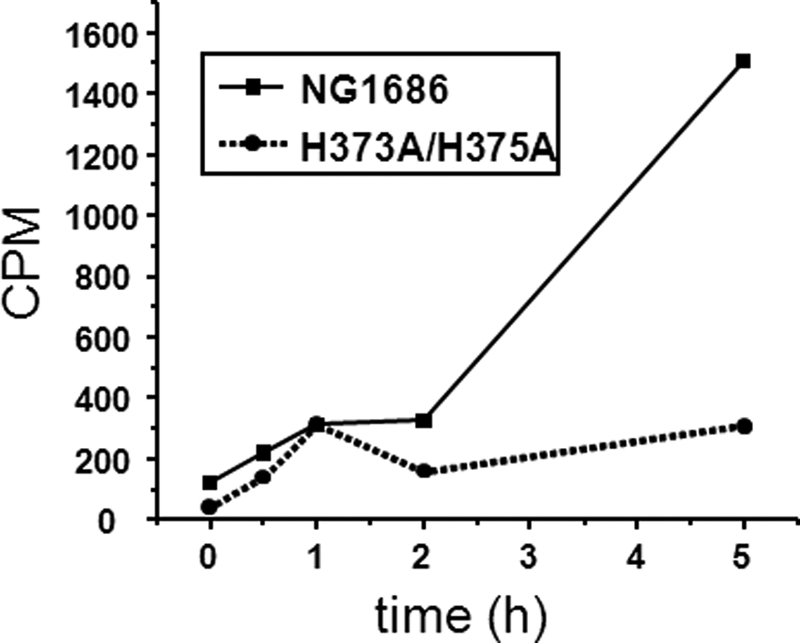
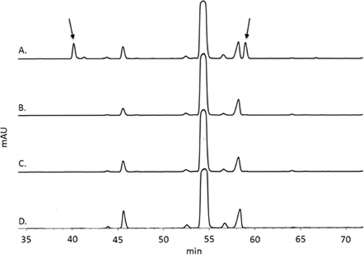
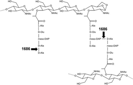
Symptomatic gonococcal infection, caused exclusively by the human-specific pathogen Neisseria gonorrhoeae (the gonococcus), is characterized by the influx of polymorphonuclear leukocytes (PMNs) to the site of infection. Although PMNs possess a potent antimicrobial arsenal comprising both oxidative and non-oxidative killing mechanisms, gonococci survive this interaction, suggesting that the gonococcus has evolved many defenses against PMN killing. We previously identified the NG1686 protein as a gonococcal virulence factor that protects against both non-oxidative PMN-mediated killing and oxidative killing by hydrogen peroxide. In this work, we show that deletion of ng1686 affects gonococcal colony morphology but not cell morphology and that overexpression of ng1686 does not confer enhanced survival to hydrogen peroxide on gonococci. NG1686 contains M23B endopeptidase active sites found in proteins that cleave bacterial cell wall peptidoglycan. Strains of N. gonorrhoeae expressing mutant NG1686 proteins with substitutions in many, but not all, conserved metallopeptidase active sites recapitulated the hydrogen peroxide sensitivity and altered colony morphology of the Δng1686 mutant strain. We showed that purified NG1686 protein degrades peptidoglycan in vitro and that mutations in many conserved active site residues abolished its degradative activity. Finally, we demonstrated that NG1686 possesses both dd-carboxypeptidase and endopeptidase activities. We conclude that the NG1686 protein is a M23B peptidase with dual activities that targets the cell wall to affect colony morphology and resistance to hydrogen peroxide and PMN-mediated killing.


Figures

FIGURE 1.
FIGURE 1.
Western blot analysis of NG1686 protein expression in parent, mutant, and complement strains. SDS-polyacrylamide gels containing 10 μg of total cellular protein per lane of strains FA1090, Δ1686 (Δng1686), and Δ1686/1686+ were run and transferred to PVDF membrane, with subsequent Western blot analysis using anti-1686 antiserum in the ECL Plus detection kit.
FIGURE 2.
FIGURE 2.
NG1686 protein expression in and H2O2 resistance of IPTG-regulatable ng1686 strains. A, representative Western blot analysis of NG1686 protein expression in strains containing IPTG-regulatable ng1686 construct pKH35/Plac1686. SDS-polyacrylamide gels containing 10 μg of total protein per lane were run, transferred, and developed as described in the legend to Fig. 1. B, dose-response curve of H2O2 resistance after 15 min of strains containing pKH35/1686. Cells were treated with varying doses of H2O2 for 15 min and serially diluted into medium containing catalase. The relative survival at each dose was calculated as viable cfu divided by total cfu (receiving no H2O2). Error bars, S.E. of 2–4 independent experiments. Strain Δ1686nv/Plac1686 (designated Δ1686nv/1686, in the absence of IPTG (−IPTG) is statistically the same as strain Δ1686nv at 20 and 50 mm H2O2 doses (*, p > 0.05) and statistically different from strain FA1090nv (†, p < 0.04) at the same doses by Student's t test. Indicated strains are statistically the same as strain FA1090nv at all doses (‡, p > 0.05).
FIGURE 3.
FIGURE 3.
Phenotypes of M23B metallopeptidase active site point mutants of NG1686 expressed in N. gonorrhoeae. A, colony morphology of N. gonorrhoeae strains containing M23B point mutants. Representative stereomicroscope observations of strains using a Nikon SMZ-10A stereomicroscope after 24 h of growth on solid medium were recorded using a Polaroid camera model DMC Ie. B, dose-response curve of H2O2 resistance after 15 min of stains carrying NG1686 M23B point mutants. Cells were treated with varying doses of H2O2 for 15 min and serially diluted into medium containing catalase. Error bars, S.E. of six independent experiments. Strains are statistically the same as strain Δ1686 at 20 and 50 mm H2O2 doses (*, p > 0.05) and statistically the same as strain Δ1686/1686+ at all H2O2 doses (†, p > 0.05) by Student's t test. C, Western blot analysis of NG1686 M23B point mutant protein expression. SDS-polyacrylamide gels containing 10 μg of total cellular protein per lane were run and transferred to PVDF membrane. Lane 1, Δ1686/1686+; lane 2, H295A; lane 3, D299A; lane 4, H295A/D299A; lane 5, H373A; lane 6, H375A; lane 7, H373A/H375A.
FIGURE 4.
FIGURE 4.
Zymogram analysis of peptidoglycan hydrolase activity. A, equal amounts of E. coli cell extract carrying pET28a/NG1686 or pET28a/H373A/H375A or purified NG1686 protein were separated by SDS-PAGE, renatured in the E. coli PG-containing gel, and stained with methylene blue. Zones of clearing corresponding to either lysozyme or NG1686 protein represent PG degradation and are indicated with arrows. B, gel subsequently stained with Coomassie shows equivalent amounts of purified NG1686 and NG1686 proteins derived from E. coli cell lysates were loaded on the gel. C, equal amounts (5 μl) of E. coli cell extracts carrying NG1686 M23B point mutant constructs were run on an N. gonorrhoeae PG-containing gel and stained with methylene blue. Zones of clearing are indicated with arrows. D, differing amounts of E. coli cell extracts carrying NG1686 M23B point mutant constructs, indicated at the bottom of E, were run on an E. coli PG-containing gel and stained with methylene blue. E, gel from D subsequently stained with Coomassie to show the relative amounts of NG1686 mutant proteins loaded on gel.
FIGURE 5.
FIGURE 5.
Degradation of radiolabeled PG by purified NG1686 and H373A/H375A proteins. Gonococcal PG, NG1686 protein, and H373A/H375A protein were incubated together. Samples of the reactions were collected at the indicated times, and the insoluble, macromolecular PG was precipitated by the addition of TCA and collected by centrifugation. The soluble fragments were quantified by scintillation counting. Values are the average of two independent experiments.
FIGURE 6.
FIGURE 6.
LC/MS characterization of soluble reaction products produced by digestion of PG by purified NG1686 protein. Base peak chromatogram showing soluble reaction products from the NG1686 enzymatic reaction (mass range, 800–3900 Da). See Table 1 for a description of the numbered peaks. These ions were not detected in control reactions containing NG1686 and EDTA (1 mm) or the H373A/H375A mutant protein. cps, counts per second.
FIGURE 7.
FIGURE 7.
HPLC analysis of FDNB-derivatized reaction mixtures containing NG1686 (A), H373A/H375A (B), or NG1686 + EDTA (C) and NG1686 reaction lacking PG (D). The arrows indicate elution products that were collected, dried, and analyzed by ESI-MS. Based on ESI-MS and the elution of standards, the first arrow corresponds to the mono-DNP-DAP; the second arrow corresponds to DNP-alanine. mAU, milliabsorbance units.
FIGURE 8.
FIGURE 8.
HPLC analysis of FDNB-derivatized reaction mixtures containing d-alanine (A), MurNAc-pentapeptide + NG1686 (B), or MurNAc-pentapeptide + H373A/H375A (C) and NG1686 reaction lacking substrate (D). The arrows indicate the elution products corresponding to DNP-alanine. mAU, milliabsorbance units.
FIGURE 9.
FIGURE 9.
Sites of PG cleavage by NG1686. Shown is a schematic representation of sites cleaved by the endopeptidase and dd-carboxypeptidase activities of NG1686.

Similar articles

Cited by

References

    1. World Health Organization (2011) Emergence of multi-drug resistant Neisseria gonorrhoeae. Threat of global rise in untreatable sexually transmitted disease. in WHO/RHR/11.4 Department of Reproductive Health and Research, World Health Organization, Geneva
    1. Hook E. W., 3rd, Holmes K. K. (1985) Gonococcal infections. Ann. Int. Med. 102, 229–243 - PubMed
    1. Edwards J. L., Apicella M. A. (2004) The molecular mechanisms used by Neisseria gonorrhoeae to initiate infection differ between men and women. Clin. Microbiol. Rev. 17, 965–981 - PMC - PubMed
    1. Dokter W. H., Dijkstra A. J., Koopmans S. B., Stulp B. K., Keck W., Halie M. R., Vellenga E. (1994) G(Anh)MTetra, a natural bacterial cell wall breakdown product, induces interleukin-1 β and interleukin-6 expression in human monocytes. A study of the molecular mechanisms involved in inflammatory cytokine expression. J. Biol. Chem. 269, 4201–4206 - PubMed
    1. Melly M. A., McGee Z. A., Rosenthal R. S. (1984) Ability of monomeric peptidoglycan fragments from Neisseria gonorrhoeae to damage human fallopian-tube mucosa. J. Infect. Dis. 149, 378–386 - PubMed

Publication types

MeSH terms