. 2012 Mar 30;287(14):11489-97.
doi: 10.1074/jbc.M111.313056. Epub 2012 Feb 13.

Successful prediction of substrate-binding pocket in SLC17 transporter sialin

Affiliations

Successful prediction of substrate-binding pocket in SLC17 transporter sialin

Nicolas Pietrancosta et al. J Biol Chem. .

Abstract

Secondary active transporters from the SLC17 protein family are required for excitatory and purinergic synaptic transmission, sialic acid metabolism, and renal function, and several members are associated with inherited neurological or metabolic diseases. However, molecular tools to investigate their function or correct their genetic defects are limited or absent. Using structure-activity, homology modeling, molecular docking, and mutagenesis studies, we have located the substrate-binding site of sialin (SLC17A5), a lysosomal sialic acid exporter also recently implicated in exocytotic release of aspartate. Human sialin is defective in two inherited sialic acid storage diseases and is responsible for metabolic incorporation of the dietary nonhuman sialic acid N-glycolylneuraminic acid. We built cytosol-open and lumen-open three-dimensional models of sialin based on weak, but significant, sequence similarity with the glycerol-3-phosphate and fucose permeases from Escherichia coli, respectively. Molecular docking of 31 synthetic sialic acid analogues to both models was consistent with inhibition studies. Narrowing the sialic acid-binding site in the cytosol-open state by two phenylalanine to tyrosine mutations abrogated recognition of the most active analogue without impairing neuraminic acid transport. Moreover, a pilot virtual high-throughput screening of the cytosol-open model could identify a pseudopeptide competitive inhibitor showing >100-fold higher affinity than the natural substrate. This validated model of human sialin and sialin-guided models of other SLC17 transporters should pave the way for the identification of inhibitors, glycoengineering tools, pharmacological chaperones, and fluorescent false neurotransmitters targeted to these proteins.


Figures
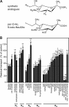
FIGURE 1.
FIGURE 1.
Structure-activity relationship of human sialin. A, a generic synthetic sialic acid analogue (top structure) and the most bioactive analogue, per-O-Ac,9-iodo-Neu5Ac, are shown under their β anomeric state. The nine carbon atoms of the Neu5Ac scaffold are numbered in italics. B, inhibition of [3H]Neu5Ac uptake by synthetic analogues (1 mm) at pH 5.0. The dotted line shows inhibition by unlabeled Neu5Ac. The numbers and asterisks above the bars indicate numbers of independent experiments and the statistical significance (Mann-Whitney test). *, p < 0.05; **, p < 0.001.
FIGURE 2.
FIGURE 2.
Molecular docking of sialic acid analogues to lumen-open and cytosol-open sialin models. Synthetic analogues were docked in silico to the three-dimensional models under their α or β anomeric state. Residual transport levels shown in Fig. 1B were plotted against the global energy of the most stable ligand-protein pose for each compound. The horizontal gray line corresponds to a 5-fold increase in affinity relative to Neu5Ac. The vertical line was positioned to separate top-scoring docked compounds from medium- and low-scoring compounds. Quadrants a, b, c, and d correspond to false negative, true negative, false positive, and true positive hits, respectively.
FIGURE 3.
FIGURE 3.
Structural snapshots of β anomer of per-O-Ac,9-iodo-Neu5Ac docked into its binding pocket. Lumen-open and cytosol-open sialin models are shown in the top and bottom rows, respectively. Left images are side views, with the lysosomal lumen (equivalent to the extracellular compartment in our transport assay) at the top. Green and blue continuous lines indicate luminal and cytosolic boundaries of the membrane, respectively. Right images are views from the cytosol. For clarity, only residues having polar interactions with the ligand (pink side chains) are displayed, along with two phenylalanine residues (green) subjected to mutagenesis in Fig. 4. Dotted green lines indicate hydrogen bonds.
FIGURE 4.
FIGURE 4.
Narrowing sialic acid-binding pocket of sialin by conservative missense mutations selectively impairs recognition of synthetic analogue per-O-Ac,9-iodo-Neu5Ac. A, space-filling models of the docked analogue (yellow) and of Phe-50 and Phe-410 side chains (green) show that the two phenylalanines make van der Waals contacts with the ligand in the cytosol-open state of human sialin. This holds true for both α and β anomers despite their distinct positions in the binding pocket. B, single and double missense mutants of Phe-50 and Phe-410 show substantial Neu5Ac transport activity. The number above the bars indicate numbers of independent experiments. C, dose-response inhibition curves of wild-type (black) and mutant (red) sialin by Neu5Ac (circles) and the synthetic analogue (triangles). Representative experiments are shown. See Table 2 for average results from independent experiments.
FIGURE 5.
FIGURE 5.
Successful identification of a novel sialin ligand by virtual high-throughput screening. A, chemical structure of the virtual hit. B, structural snapshots of FR139317 docked into its binding pocket of the cytosol-open model. Data are displayed as in Fig. 3. Most side chains interacting with the pseudopeptide are also involved in sialic acid analogue binding. C, representative dose-response inhibition curve of FR139317. D, FR139317 competitively inhibits Neu5Ac uptake. A representative saturation kinetics experiment is shown as an Eadie-Hofstee plot. Where not visible, error bars are smaller than symbols. E, the apparent Michaelis-Menten constant (Kmapp) derived from regression lines in D is plotted as a function of the FR139317 concentration. Kmapp increases linearly with this concentration (r2 = 0.9776), yielding an inhibitory constant of 9.0 μm in this experiment.

References

    1. Law C. J., Maloney P. C., Wang D. N. (2008) Ins and outs of major facilitator superfamily antiporters. Annu. Rev. Microbiol. 62, 289–305 - PMC - PubMed
    1. Pao S. S., Paulsen I. T., Saier M. H., Jr. (1998) Major facilitator superfamily. Microbiol. Mol. Biol. Rev. 62, 1–34 - PMC - PubMed
    1. Abramson J., Smirnova I., Kasho V., Verner G., Kaback H. R., Iwata S. (2003) Structure and mechanism of the lactose permease of Escherichia coli. Science 301, 610–615 - PubMed
    1. Dang S., Sun L., Huang Y., Lu F., Liu Y., Gong H., Wang J., Yan N. (2010) Structure of a fucose transporter in an outward-open conformation. Nature 467, 734–738 - PubMed
    1. Reimer R. J., Edwards R. H. (2004) Organic anion transport is the primary function of the SLC17/type I phosphate transporter family. Pflugers Arch. 447, 629–635 - PubMed

Publication types

MeSH terms

LinkOut - more resources