Review
. 2012 Apr;12(2):110-28.
doi: 10.2174/187152612800100143.

Targeting RSV with vaccines and small molecule drugs

Affiliations
Review

Targeting RSV with vaccines and small molecule drugs

Heather M Costello et al. Infect Disord Drug Targets. 2012 Apr.

Abstract

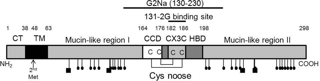
Respiratory syncytial virus (RSV) is the most significant cause of pediatric respiratory infections. Palivizumab (Synagis®), a humanized monoclonal antibody, has been used successfully for a number of years to prevent severe RSV disease in at-risk infants. However, despite intense efforts, there is no approved vaccine or small molecule drug for RSV. As an enveloped virus, RSV must fuse its envelope with the host cell membrane, which is accomplished through the actions of the fusion (F) glycoprotein, with attachment help from the G glycoprotein. Because of their integral role in initiation of infection and their accessibility outside the lipid bilayer, these proteins have been popular targets in the discovery and development of antiviral compounds and vaccines against RSV. This review examines advances in the development of antiviral compounds and vaccine candidates.


Figures

Figure 1
Figure 1
Monomer model of the pretriggered (A) RSV F protein based on the crystal structure of the pretriggered PIV5 F protein, and the crystal structure of the posttriggered RSV F protein (B) (PDB structure 3RRR) [57, 58]. We have maintained the HRB domain (red α-helix) in approximately the same position in both models, as a fixed reference point. Following triggering, the pre-HRA domain (blue in A) becomes one long α-helix (blue in B), inserts its N-terminal fusion peptide (white in A) into the target membrane and folds back (B), bringing the virion and cell membranes together. The fusion peptide was not resolved in the posttriggered structure. During this fold-back, the “Head” domain maintains its position relative to HRA, but rotates approximately 180 degrees, bottom to top, relative to HRB. This rotation causes the peptide that connects HRB to the Head to wind off the Head. Two of the three N-linked glycosylation sites are indicated. The third N-linked site would be at the N-terminus of F2 (on the surface of the “Head” region) if the preceding amino acid of F2 had been visualized in the PIV5 F protein pretriggered structure. Cysteines, all of which are involved in disulfide bonds appear as yellow balls. Drug-resistant mutations appear as red balls. Residues 140 and 144 are not present in the posttriggered structure, because this region of the fusion peptide (the 9 N-terminal residues) were deleted from the nucleotide sequence to prevent aggregation of the posttriggered form. In addition, the fusion peptide was not resolved. Residue Y198 is presented as blue spheres.
Figure 2
Figure 2
Cartoon of dynamic regions of the F protein that refold to initiate fusion. The three N-terminal α-helices in pre-HRA are connected by two non-helical peptides in the pretriggered form. Upon triggering these non-helical connecting peptides refold into α-helices, completing the long HRA α-helix (dark gray) and in the process thrust the F1 N-terminal fusion peptide into the target cell membrane. HRA trimerizes, the molecule folds in half, and the HRB α-helices (light gray) insert into the grooves on the surface of the HRA trimer forming the stable 6-helix bundle (6HB). As a result, the virion and cell membranes are brought together and initiate membrane fusion. The “head” region of the F protein does not rearrange during the triggering and refolding events and, therefore, is not represented in this cartoon.
Figure 3
Figure 3
Cartoon of the 298 amino acid long RSV G protein showing the N-terminal location of its cytoplasmic domain (CD) and transmembrane domain (TM), two mucin-like regions with their N-linked (squares) and O-linked (small ovals) glycans, and central cysteine noose which contains the 4 conserved cysteines, the central conserved domain (CCD), and the CX3C domain, followed by the heparin-binding domain (HBD). The position of the mAb 131-2G binding site and the peptide included in the experimental G2Na vaccine are indicated. The position of the second Met that is used to initiate translation of a truncated form of the G protein is also indicated. This form is proteolytically cleaved to remove its truncated TM domain and released from the cell as soluble G protein.

References

    1. Collins PLaJEC., Jr . Respiratory syncytial virus and metapneumovirus. Philadelphia: Lippincott Williams & Wilkins; 2007.
    1. Glezen P, Denny FW. Epidemiology of acute lower respiratory disease in children. N Engl J Med. 1973;288(10):498–505. - PubMed
    1. Kim HW, Arrobio JO, Brandt CD, Jeffries BC, Pyles G, Reid JL, Chanock RM, Parrott RH. Epidemiology of respiratory syncytial virus infection in Washington, D.C. I. Importance of the virus in different respiratory tract disease syndromes and temporal distribution of infection. Am J Epidemiol. 1973;98(3):216–225. - PubMed
    1. Ogra PL, Patel J. Respiratory syncytial virus infection and the immunocompromised host. Pediatr Infect Dis J. 1988;7(4):246–249. - PubMed
    1. Pohl C, Green M, Wald ER, Ledesma-Medina J. Respiratory syncytial virus infections in pediatric liver transplant recipients. J Infect Dis. 1992;165(1):166–169. - PubMed

Publication types

MeSH terms

Substances