. 2012 Apr 10;29(6):1233-42.
doi: 10.1089/neu.2011.2169. Epub 2012 Apr 10.

Challenging the role of adaptive immunity in neurotrauma: Rag1(-/-) mice lacking mature B and T cells do not show neuroprotection after closed head injury

Affiliations

Challenging the role of adaptive immunity in neurotrauma: Rag1(-/-) mice lacking mature B and T cells do not show neuroprotection after closed head injury

Sebastian Weckbach et al. J Neurotrauma. .

Abstract

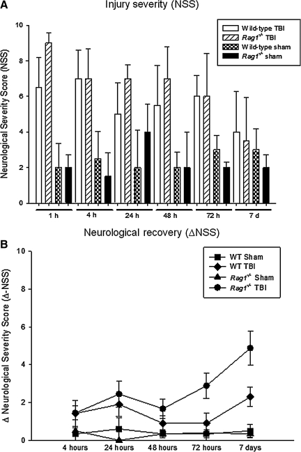
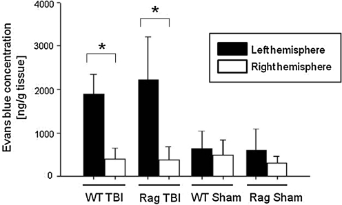
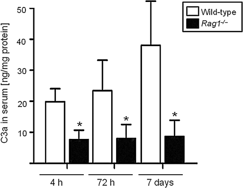
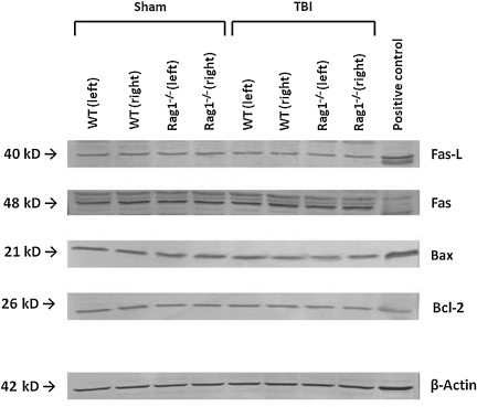
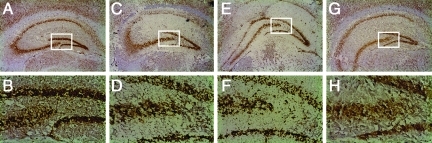
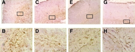
The role of adaptive immunity in contributing to post-traumatic neuroinflammation and neuropathology after head injury remains largely unexplored. The present study was designed to investigate the pathophysiological sequelae of closed head injury in Rag1(-/-) mice devoid of mature B and T lymphocytes. C57BL/6 wild-type and Rag1(-/-) mice were subjected to experimental closed head injury, using a standardized weight-drop device. Outcome parameters consisted of neurological scoring, quantification of blood-brain barrier (BBB) function, measurement of inflammatory markers and mediators of apoptosis in serum and brain tissue, and assessment of neuronal cell death, astrogliosis, and tissue destruction. There was no difference between wild-type and Rag1(-/-) mice with regard to injury severity and neurological impairment for up to 7 days after head injury. The extent of BBB dysfunction was in a similar range for both groups. Quantification of complement activation fragments in serum revealed significantly attenuated C3a levels in Rag1(-/-) mice compared to wild-type animals. In contrast, the levels of pro- and anti-inflammatory cytokines and pro-apoptotic and anti-apoptotic mediators remained in a similar range for both groups, and the histological analysis of brain sections did not reveal a difference in reactive astrogliosis, tissue destruction, and neuronal cell death in Rag1(-/-) compared to wild-type mice. These findings suggest that adaptive immunity is not of crucial importance for initiating and sustaining the inflammatory neuropathology after closed head injury. The attenuated extent of post-traumatic complement activation seen in Rag1(-/-) mice implies a cross-talk between innate and adaptive immune responses, which requires further investigation in future studies.


Figures

FIG. 1.
FIG. 1.
Assessment of injury severity, neurological impairment, and recovery in C57BL/6 wild-type (n=97) and Rag1−/− mice (n=87) subjected to closed head injury or sham surgery, using a standardized 10-point Neurological Severity Score (NSS; A). A maximal score of 10 points corresponds to severe neurological impairment, whereas a low score of 0–3 points reflects normal physiological behavior. The ΔNSS (B), calculated as the difference between the NSS at 1 h and the NSS at any later time point, represents a parameter reflecting the degree of spontaneous recovery after TBI, as previously described. All data are presented as medians±standard deviation. No statistically significant differences were seen in NSS between head-injured wild-type and Rag1−/− mice at any time point assessed. In contrast, head-injured Rag1−/− mice showed improved recovery by 7 days, as reflected by a significantly increased ΔNSS at 7 days, compared to wild-type animals (p<0.01; TBI, traumatic brain injury).
FIG. 2.
FIG. 2.
Quantification of post-traumatic blood–brain barrier dysfunction in C57BL/6 wild-type (n=11) and Rag1−/− mice (n=14) at 4 h after closed head injury or sham surgery. Evans blue extravasation into the injured (left) and non-injured (right) brain hemispheres was quantified by fluorospectrophotometry, as outlined in the methods section. Data are presented as medians±standard deviation; (*p<0.05 for left versus right hemisphere in head-injured mice, and for left hemisphere in head-injured versus sham-operated mice in both groups; TBI, traumatic brain injury).
FIG. 3.
FIG. 3.
Complement anaphylatoxin C3a serum concentrations in C57BL/6 wild-type (n=22) and Rag1−/− mice (n=23) at 4 h, 72 h, and 7 days after closed head injury. C3a levels were determined by a mouse-specific ELISA and normalized by total protein concentration, as described in the methods section. Data are shown as medians±standard deviation (*p<0.05 for wild-type versus Rag1−/− mice at all time points; ELISA, enzyme-linked immunosorbent assay).
FIG. 4.
FIG. 4.
Representative Western blot analysis of brain homogenates from injured (left) and uninjured (right) hemispheres of C57BL/6 wild-type (WT) and Rag1−/− mice at 4 h after sham surgery or closed head injury. Equal concentrations of protein (50 μg per lane) were loaded on SDS-Page membranes, and consistent loading was confirmed by β-actin control blotting. Mouse-specific primary antibodies against Fas, FasL, Bax, and Bcl-2, were used on nitrocellulose membranes and visualized by a colorimetric assay using alkaline phosphatase, as described in the methods section.
FIG. 5.
FIG. 5.
Neuronal morphology and microarchitecture in the hippocampus of sham-operated (AD) and head-injured (EH) wild-type and Rag1−/− mice, as determined by immunohistochemistry in 10-μm-thick coronal brain cryosections. A monoclonal anti-NeuN antibody was used as a neuron-specific marker. Adult male C57BL/6 and Rag1−/− mice (n=3 per group) were euthanized at 24 h after the surgical procedure. The lower panels represent fivefold magnifications of the boxed areas in their respective upper panels (100×and 20×original magnifications), depicting the hippocampal CA3/CA4 cell layers. Sham-operated wild-type (A and B) and Rag1−/− mice (C and D) showed a similar hippocampal structure and neuronal morphology. In contrast, head injury induced cellular disruption and changes in neuronal morphology in the CA3/CA4 cell layers; however, this did not differ in extent between wild-type (E and F) and Rag1−/− mice (G and H).
FIG. 6.
FIG. 6.
Astroglial reaction in injured mouse brains at 7 days after closed head injury. Immunohistochemical staining of 10-μm-thick coronal brain was done using glial fibrillary acidic protein (GFAP) as a specific cell marker for astrocytes. Sham-operated wild-type (A and B), and Rag1−/− mice (C and D), showed a similar baseline morphology of GFAP expression, with the classic feature of non-reactive astrocytes. In contrast, closed head injury induced reactive astrocytosis in the injured cortex of wild-type (E and F) and Rag1−/− mice (G and H), with identical changes in morphology in both mouse strains by 7 days after trauma. The lower panels represent fivefold magnifications of the boxed areas of their respective upper panels (100×and 20×original magnifications).

Similar articles

Cited by

References

    1. Alexander J.J. Anderson A.J. Barnum S.R. Stevens B. Tenner A.J. The complement cascade: Yin-Yang in neuroinflammation, neuroprotection, and degeneration. J. Neurochem. 2008;107:1169–1187. - PMC - PubMed
    1. Ankeny D.P. Guan Z. Popovich P.G. B cells produce pathogenic antibodies and impair recovery after spinal cord injury in mice. J. Clin. Invest. 2009;119:2990–2999. - PMC - PubMed
    1. Austen W.G., Jr. Zhang M. Chan R. Friend D. Hechtman H.B. Carroll M.C. Moore F.D., Jr. Murine hindlimb reperfusion injury can be initiated by a self-reactive monoclonal IgM. Surgery. 2004;136:401–406. - PubMed
    1. Beauchamp K. Mutlak H. Smith W.R. Shohami E. Stahel P.F. Pharmacology of traumatic brain injury: where is the “golden bullet”? Mol. Med. 2008;14:731–740. - PMC - PubMed
    1. Bechmann I. Galea I. Perry V.H. What is the blood-brain barrier (not)? Trends Immunol. 2007;28:5–11. - PubMed

Publication types

LinkOut - more resources