Review
. 2012 Aug;18(4):288-300.
doi: 10.1089/ten.TEB.2011.0465. Epub 2012 Mar 28.

Mechanical stretching for tissue engineering: two-dimensional and three-dimensional constructs

Affiliations
Review

Mechanical stretching for tissue engineering: two-dimensional and three-dimensional constructs

Brandon D Riehl et al. Tissue Eng Part B Rev. 2012 Aug.

Abstract

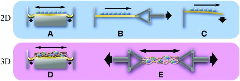
Mechanical cell stretching may be an attractive strategy for the tissue engineering of mechanically functional tissues. It has been demonstrated that cell growth and differentiation can be guided by cell stretch with minimal help from soluble factors and engineered tissues that are mechanically stretched in bioreactors may have superior organization, functionality, and strength compared with unstretched counterparts. This review explores recent studies on cell stretching in both two-dimensional (2D) and three-dimensional (3D) setups focusing on the applications of stretch stimulation as a tool for controlling cell orientation, growth, gene expression, lineage commitment, and differentiation and for achieving successful tissue engineering of mechanically functional tissues, including cardiac, muscle, vasculature, ligament, tendon, bone, and so on. Custom stretching devices and lab-specific mechanical bioreactors are described with a discussion on capabilities and limitations. While stretch mechanotransduction pathways have been examined using 2D stretch, studying such pathways in physiologically relevant 3D environments may be required to understand how cells direct tissue development under stretch. Cell stretch study using 3D milieus may also help to develop tissue-specific stretch regimens optimized with biochemical feedback, which once developed will provide optimal tissue engineering protocols.


Figures

FIG. 1.
FIG. 1.
Schematic of 2D and 3D stretching devices. Stretching of a 2D cell layer is achieved via pneumatic deformation of the membrane (A), clamp arm mechanism (B), and bending (C). Stretching of 3D cell–scaffold constructs is accomplished via pneumatic deformation (D) and clamping mechanism (E). 2D, two-dimensional; 3D, three-dimensional. Color images available online at www.liebertonline.com/teb
FIG. 2.
FIG. 2.
Cell stretching in 2D affects cell orientation and cytoskeletal development, showing directional dependency. C2C12 myoblasts were cyclically stretched at 7% and 0.5 Hz in horizontal direction on membranes with micropatterned fibronectin lines. Staining with actin is shown. (a) Unpatterned (homogeneous [HS]), unstretched control had less well-developed actin fibers. (b) Stretching of unpatterned cells developed actin stress fibers oriented at an average angle of 72° (cyclic tensile strain [CTS]). (c) Patterned but not stretched cells showed actin fibers oriented along the fibronectin line pattern. (d) Stretch applied parallel to the patterned cell direction induced irregular cell orientation and formed actin stress fibers oblique to the strain direction (average actin fiber orientation of 48°). (e) Stretch applied to 45° of patterns caused an average actin fiber orientation of 52°. (f) Perpendicular stretch induced actin fiber orientation of 91°. Scale bar=50 μm. Reprinted with permission from Elsevier (Ahmed et al.).
FIG. 3.
FIG. 3.
Cell stretching in 3D affects actin stress fiber formation depending on static or dynamic stretching condition. Rat bone-marrow-derived progenitor cells were cultured in fibrin scaffolds. (A) Free-float, unstretched control showed random cell orientation. (B) Static stress group (constrained between anchors but not stretched) oriented actin fibers parallel to the direction of strain (arrows). (C) Cyclic stretch (10%, 1 Hz, for 6 days) oriented actin fibers parallel to the strain direction (arrows). (D) Quantified stress filament area per cell shows an increase in the order of free float<static constrained<dynamic stretch. Blue is DAPI staining and green is F-actin staining. Scale bar=10 μm (insets are at 100× magnification). Reprinted with permission from Wiley (Nieponice et al.). Color images available online at www.liebertonline.com/teb
FIG. 4.
FIG. 4.
Major stretch-induced mechanotransduction signaling elements, including integrins, force-sensitive kinases and proteins, cytoskeletal elements, stretch-activated ion channels, membranes, ions, and so on. ERK, extracellular-signal-regulated kinase; FAK, focal adhesion kinase; MAPK, mitogen-activated protein kinase; Pax, paxillin; ROCK, RhoA kinase; Tal, talin; TGF, transforming growth factor; VASP, vasodilator-stimulated phosphoprotein; VEGF, vascular endothelial growth factor; Vin, vinculin. Color images available online at www.liebertonline.com/teb

References

    1. Powell H.M. McFarland K.L. Butler D.L. Supp D.M. Boyce S.T. Uniaxial strain regulates morphogenesis, gene expression, and tissue strength in engineered skin. Tissue Eng Part A. 2010;16:1083. - PubMed
    1. Lee Y.U. Hayman D. Sprague E.A. Han H.C. Effects of axial stretch on cell proliferation and intimal thickness in arteries in organ culture. Cell Mol Bioeng. 2010;3:286. - PMC - PubMed
    1. Benhardt H.A. Cosgriff-Hernandez E.M. The role of mechanical loading in ligament tissue engineering. Tissue Eng Part B Rev. 2009;15:467. - PubMed
    1. Lee W.C. Maul T.M. Vorp D.A. Rubin J.P. Marra K.G. Effects of uniaxial cyclic strain on adipose-derived stem cell morphology, proliferation, and differentiation. Biomech Model Mechanobiol. 2007;6:265. - PubMed
    1. Kelly D.J. Jacobs C.R. The role of mechanical signals in regulating chondrogenesis and osteogenesis of mesenchymal stem cells. Birth Defects Res C Embryo Today. 2010;90:75. - PubMed

Publication types

LinkOut - more resources