

Assessment of left ventricular ejection fraction using an ultrasonic stethoscope in critically ill patients
- PMID: 22335818
- PMCID: PMC3396274
- DOI: 10.1186/cc11198
Assessment of left ventricular ejection fraction using an ultrasonic stethoscope in critically ill patients
Abstract
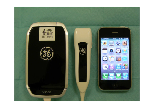
Introduction: Assessment of cardiac function is key in the management of intensive care unit (ICU) patients and frequently relies on the use of standard transthoracic echocardiography (TTE). A commercially available new generation ultrasound system with two-dimensional imaging capability, which has roughly the size of a mobile phone, is adequately suited to extend the physical examination. The primary endpoint of this study was to evaluate the additional value of this new miniaturized device used as an ultrasonic stethoscope (US) for the determination of left ventricular (LV) systolic function, when compared to conventional clinical assessment by experienced intensivists. The secondary endpoint was to validate the US against TTE for the semi-quantitative assessment of left ventricular ejection fraction (LVEF) in ICU patients.
Methods: In this single-center prospective descriptive study, LVEF was independently assessed clinically by the attending physician and echocardiographically by two experienced intensivists trained in critical care echocardiography who used the US (size: 135×73×28 mm; weight: 390 g) and TTE. LVEF was visually estimated semi-quantitatively and classified in one of the following categories: increased (LVEF>75%), normal (LVEF: 50 to 75%), moderately reduced (LVEF: 30 to 49%), or severely reduced (LVEF<30%). Biplane LVEF measured using the Simpson's rule on TTE loops by an independent investigator was used as reference.
Results: A total of 94 consecutive patients were studied (age: 60±17 years; simplified acute physiologic score 2: 41±15), 63 being mechanically ventilated and 36 receiving vasopressors and/or inotropes. Diagnostic concordance between the clinically estimated LVEF and biplane LVEF was poor (Kappa: 0.33; 95% CI: 0.16 to 0.49) and only slightly improved by the knowledge of a previously determined LVEF value (Kappa: 0.44; 95% CI: 0.22 to 0.66). In contrast, the diagnostic agreement was good between visually assessed LVEF using the US and TTE (Kappa: 0.75; CI 95%: 0.63 to 0.87) and between LVEF assessed on-line and biplane LVEF, regardless of the system used (Kappa: 0.75; CI 95%: 0.64 to 0.87 and Kappa: 0.70; CI 95%: 0.59 to 0.82, respectively).
Conclusions: In ICU patients, the extension of physical examination using an US improves the ability of trained intensivists to determine LVEF at bedside. With trained operators, the semi-quantitative assessment of LVEF using the US is accurate when compared to standard TTE.
Figures
References
-
- Iregui MG, Prentice D, Sherman G, Schallom L, Sona C, Kollef MH. Physicians' estimates of cardiac index and intravascular volume based on clinical assessment versus transesophageal Doppler measurements obtained by critical care nurses. Am J Crit Care. 2003;12:336–342. - PubMed
-
- Task Force for Diagnosis and Treatment of Acute and Chronic Heart Failure 2008 of European Society of Cardiology; Dickstein K, Cohen-Solal A, Filippatos G, McMurray JJ, Ponikowski P, Poole-Wilson PA, Strömbert A, van Veldhuisen DJ, Atar D, Hoes AW, Keren A, Mebazaa A, Nieminen M, Priori SG, Swedberg K. ESC Committee for Practice Guidelines. Vahanian A, Camm J, De Caterina R, Dean V, Dickstein K, Filippatos G, Funck-Brentano C, Hellemans I, Kristensen SD, McGregor K, Sechtem U, Silber S, Tendera M, Widimsky P, Zamorano JL. ESC Guidelines for the diagnosis and treatment of acute and chronic heart failure 2008: the Task Force for the Diagnosis and Treatment of Acute and Chronic Heart Failure 2008 of the European Society of Cardiology. Developed in collaboration with the Heart Failure Association of the ESC (HFA) and endorsed by the European Society of Intensive Care Medicine (ESICM) Eur Heart J. 2008;29:2388–2442. - PubMed
MeSH terms
LinkOut - more resources
Full Text Sources
