

Cervical cytological changes in HIV-infected patients attending care and treatment clinic at Muhimbili National Hospital, Dar es Salaam, Tanzania
- PMID: 22335893
- PMCID: PMC3298791
- DOI: 10.1186/1750-9378-7-3
Cervical cytological changes in HIV-infected patients attending care and treatment clinic at Muhimbili National Hospital, Dar es Salaam, Tanzania
Abstract
Background: Tanzania is among Sub-Saharan countries mostly affected by the HIV and AIDS pandemic, females being more vulnerable than males. HIV infected women appear to have a higher rate of persistent infection by high risk types of human papillomavirus (HPV) strongly associated with high-grade squamous intraepithelial lesions (HSIL) and invasive cervical carcinoma. Furthermore, although HIV infection and cervical cancer are major public health problems, the frequency and HIV/HPV association of cervical cancer and HSIL is not well documented in Tanzania, thus limiting the development of preventive and therapeutic strategies.
Methods: A prospective unmatched, case-control study of HIV-seropositive, ≥ 18 years of age and consenting non-pregnant patients attending the care and treatment center (CTC) at Muhimbili National Hoospital (MNH) as cases was done between 2005 and 2006. HIV seronegative, non-pregnant and consenting women recruited from the Cervical Cancer Screening unit (CCSU) at ORCI were used as controls while those who did not consent to study participation and/or individuals under < 18 years were excluded. Pap smears were collected for routine cytodiagnosis and P53 immunohistochemistry (IHC). Cervical lesions were classified according to the Modified Bethesda System.
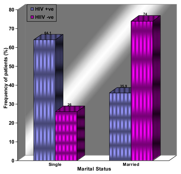
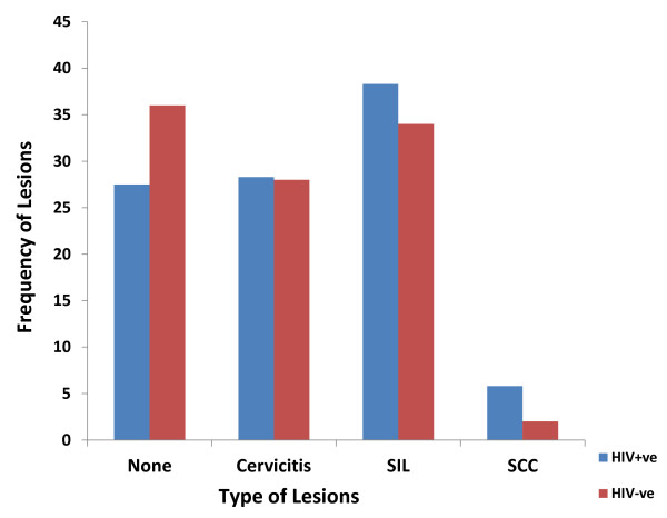
Results: A total of 170 participants from the two centers were recruited including 50 HIV-seronegative controls were from the CCSU. Ages ranged from 20-66 years (mean 40.5 years) for cases and 20-69 years (mean 41.6 years) for controls. The age group 36-45 years was the most affected by HIV (39.2%, n = 47). Cervicitis, squamous intraepithelial lesions (SIL) and carcinoma constituted 28.3% (n = 34), 38.3% (n = 46) and 5.8% (n = 7) respectively among cases, and 28% (n = 14), 34% (n = 17) and 2% (n = 1) for controls, although this was not statistically significant (P-value = 0.61). IHC showed that p53 was not detectable in HPV + Pap smears and cell blocks indicating possible degradation.
Conclusions: The frequency of SIL and carcinoma appeared to be higher among HIV-infected women on HAART compared to seronegative controls and as expected increased with age. HIV seropositive patients appeared to present earlier with SIL compared to those HIV seronegative suggesting a role of HIV in altering the natural history of HPV infection and cervical lesions. The absence of p53 immunoreactivity in HPV + lesions is indicative of the ability of HPV E6 proteins to interact with the tumor suppressor gene and pave way for viral-induced oncogenesis in the studied Tanzanian women.
Figures
Similar articles
-
Human papillomavirus (HPV) infection, HIV infection and cervical cancer in Tanzania, east Africa.Int J Cancer. 1992 Jun 19;51(4):515-21. doi: 10.1002/ijc.2910510403. Int J Cancer. 1992. PMID: 1318265
-
Epidemiology of cervical human papillomavirus (HPV) infection and squamous intraepithelial lesions (SIL) among a cohort of HIV-infected and uninfected Ghanaian women.BMC Cancer. 2017 Oct 16;17(1):688. doi: 10.1186/s12885-017-3682-x. BMC Cancer. 2017. PMID: 29037188 Free PMC article.
-
Performance of visual inspection with acetic acid and human papillomavirus testing for detection of high-grade cervical lesions in HIV positive and HIV negative Tanzanian women.Int J Cancer. 2014 Aug 15;135(4):896-904. doi: 10.1002/ijc.28712. Epub 2014 Feb 4. Int J Cancer. 2014. PMID: 24391021
-
Anal dysplasia screening: an evidence-based analysis.Ont Health Technol Assess Ser. 2007;7(4):1-43. Epub 2007 Jun 1. Ont Health Technol Assess Ser. 2007. PMID: 23074504 Free PMC article.
-
Human papillomavirus infection in women infected with the human immunodeficiency virus.N Engl J Med. 1997 Nov 6;337(19):1343-9. doi: 10.1056/NEJM199711063371903. N Engl J Med. 1997. PMID: 9358128
Cited by
-
Characteristics and geographic distribution of HIV-positive women diagnosed with cervical cancer in Dar es Salaam, Tanzania.Int J STD AIDS. 2016 Oct;27(12):1049-1056. doi: 10.1177/0956462415606252. Epub 2015 Oct 12. Int J STD AIDS. 2016. PMID: 26464500 Free PMC article.
-
Associations between highly active antiretroviral therapy and the presence of HPV, premalignant and malignant cervical lesions in sub-Saharan Africa, a systematic review: current evidence and directions for future research.BMJ Open. 2017 Aug 4;7(8):e015123. doi: 10.1136/bmjopen-2016-015123. BMJ Open. 2017. PMID: 28780541 Free PMC article.
-
High-risk HPV genotypes in Zimbabwean women with cervical cancer: Comparative analyses between HIV-negative and HIV-positive women.PLoS One. 2021 Sep 28;16(9):e0257324. doi: 10.1371/journal.pone.0257324. eCollection 2021. PLoS One. 2021. PMID: 34582476 Free PMC article.
-
Human papillomavirus genotype profiles and cytological grades interlinkages in coinfection with HIV.Pan Afr Med J. 2020 Mar 10;35:67. doi: 10.11604/pamj.2020.35.67.21539. eCollection 2020. Pan Afr Med J. 2020. PMID: 32537071 Free PMC article.
-
A Study of Pap Smear in HIV-Positive Females.J Obstet Gynaecol India. 2016 Dec;66(6):453-459. doi: 10.1007/s13224-016-0908-9. Epub 2016 Jun 6. J Obstet Gynaecol India. 2016. PMID: 27821987 Free PMC article.
References
LinkOut - more resources
Full Text Sources
Research Materials
Miscellaneous
