

Involvement of sulfoquinovosyl diacylglycerol in DNA synthesis in Synechocystis sp. PCC 6803
- PMID: 22336148
- PMCID: PMC3311599
- DOI: 10.1186/1756-0500-5-98
Involvement of sulfoquinovosyl diacylglycerol in DNA synthesis in Synechocystis sp. PCC 6803
Abstract
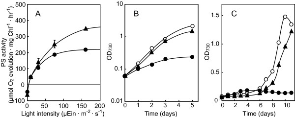
Background: Sulfoquinovosyl diacylglycerol (SQDG) is present in the membranes of cyanobacteria and their postulated progeny, plastids, in plants. A cyanobacterium, Synechocystis sp. PCC 6803, requires SQDG for growth: its mutant (SD1) with the sqdB gene for SQDG synthesis disrupted can grow with external supplementation of SQDG. However, upon removal of SQDG from the medium, its growth is retarded, with a decrease in the cellular content of SQDG throughout cell division, and finally ceases. Concomitantly with the decrease in SQDG, the maximal activity of photosynthesis at high-light intensity is repressed by 40%.
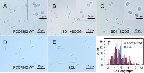
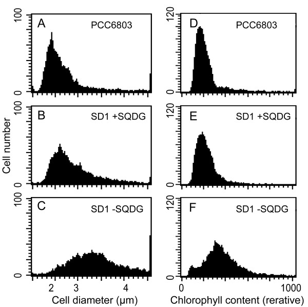
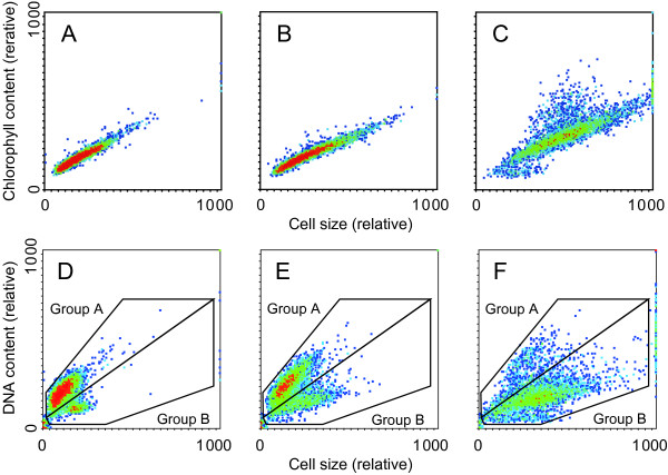
Findings: We investigated effects of SQDG-defect on physiological aspects in Synechocystis with the use of SD1. SD1 cells defective in SQDG exhibited normal photosynthesis at low-light intensity as on culturing. Meanwhile, SD1 cells defective in SQDG were impaired in light-activated heterotrophic growth as well as in photoautotrophic growth. Flow cytometric analysis of the photoautotrophically growing cells gave similar cell size histograms for the wild type and SD1 supplemented with SQDG. However, the profile of SD1 defective in SQDG changed such that large part of the cell population was increased in size. Of particular interest was the microscopic observation that the mitotic index, i.e., population of dumbbell-like cells with a septum, increased from 14 to 29% in the SD1 culture without SQDG. Flow cytometric analysis also showed that the enlarged cells of SD1 defective in SQDG contained high levels of Chl, however, the DNA content was low.
Conclusions: Our experiments strongly support the idea that photosynthesis is not the limiting factor for the growth of SD1 defective in SQDG, and that SQDG is responsible for some physiologically fundamental process common to both photoautotrophic and light-activated heterotrophic growth. Our findings suggest that the SQDG-defect allows construction of the photosynthetic machinery at an elevated level for an increase in cell mass, but represses DNA synthesis. SQDG may be essential for normal replication of chromosomal DNA for completion of the cell cycle.
© 2012 Aoki et al; BioMed Central Ltd.
Figures
References
-
- Sato N. Roles of the acidic lipids sulfoquinovosyl diacylglycerol and phosphatidylglycerol in photosynthesis: their specificity and evolution. J Plant Res. 2004;117:495–505. - PubMed
-
- Sato N, Sonoike K, Tsuzuki M, Kawaguchi A. Impaired photosystem II in a mutant of Chlamydomonas reinhardtii defective in sulfoquinovosyl diacylglycerol. Eur J Biochem. 1995;234:16–23. - PubMed
-
- Aoki M, Sato N, Meguro A, Tsuzuki M. Differing involvement of sulfoquinovosyl diacylglycerol in photosystem II in two species of unicellular cyanobacteria. Eur J Biochem. 2004;271:685–693. - PubMed
-
- Guskov A, Kern J, Gabdulkhakov A, Broser M, Zouni A, Saenger W. Cyanobacterial photosystem II at 2.9-A resolution and the role of quinones, lipids, channels and chloride. Nat Struct Mol Biol. 2009;16:334–342. - PubMed
Publication types
MeSH terms
Substances
LinkOut - more resources
Full Text Sources
