. 2012 Apr 6;287(15):11878-90.
doi: 10.1074/jbc.M111.297804. Epub 2012 Feb 15.

Rac1 protein regulates glycogen phosphorylase activation and controls interleukin (IL)-2-dependent T cell proliferation

Affiliations

Rac1 protein regulates glycogen phosphorylase activation and controls interleukin (IL)-2-dependent T cell proliferation

Onetsine Arrizabalaga et al. J Biol Chem. .

Abstract

Small GTPases of the Rho family have been implicated in important cellular processes such as cell migration and adhesion, protein secretion, and/or gene transcription. In the lymphoid system, these GTPases participate in the signaling cascades that are activated after engagement of antigen receptors. However, little is known about the role that Rho GTPases play in IL-2-mediated responses. Here, we show that IL-2 induces Rac1 activation in Kit 225 T cells. We identified by mass spectrometry the muscle isoform of glycogen phosphorylase (PYGM) as a novel Rac1 effector molecule in IL-2-stimulated cells. The interaction between the active form of Rac1 (Rac1-GTP) and PYGM was established directly through a domain comprising amino acids 191-270 of PYGM that exhibits significant homology with the Rac binding domain of PAK1. The integrity of this region was crucial for PYGM activation. Importantly, IL-2-dependent cellular proliferation was inhibited upon blocking both the activation of Rac1 and the activity of PYGM. These results reveal a new role for Rac1 in cell signaling, showing that this GTPase triggers T cell proliferation upon IL-2 stimulation by associating with PYGM and modulating its enzymatic activity.


Figures
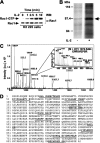
FIGURE 1.
FIGURE 1.
IL-2 leads to Rac1 activation and facilitates Rac1/PYGM association in Kit 225 cells. A, Kit 225 cells were stimulated with 500 units/ml IL-2 for several time intervals, and cell extracts were used to measure Rac1 activation. The upper panel shows the active form of Rac1 (GTP), and the lower panel shows total Rac1 in cell lysates. B, IL-2-deprived cells were stimulated with 500 units/ml IL-2 for 5 min, and protein extracts were precipitated with GST-Rac1G12V, separated by SDS-PAGE, and visualized by ProQ Diamond staining. The arrow shows the band selected for trypsin digestion. C, MALDI-TOF analysis of the excised band. Trypsin self-digestion peptides are labeled as TRYP. The peptides labeled in black matched the sequence of PYGM, and the peptides labeled in gray and underscored matched the Rac1 sequence. The inset shows the mass spectrum of MALDI-TOF/TOF (peptide fragment fingerprinting) analysis of peptide 1072.5 and its predicted amino acid sequence. D, amino acid sequence of the identified peptides covering 12% of the amino acid sequence of PYGM (GenBankTM accession number NM_005609) (shown in bold and underscored). Results are representative of three independent experiments. WB, Western blot; a.u., arbitrary units.
FIGURE 2.
FIGURE 2.
Glycogen phosphorylase is a new Rac1 GTPase-specific effector molecule. A, TaqMan gene expression assay for pygl and pygm from unstimulated and IL-2-stimulated Kit 225 cells (16 units/ml IL-2 for 24 h). ywhaz was used as a gene expression control. The results are representative of three independent experiments ± S.D. B, comparative analysis of the sequence of the putative new effector domain of PYGM with PAK1, Rhotekin, and WASP. Identical amino acids are marked by an asterisk (*), the amino acids at the same position that belong to the same strong group are marked by a colon (:), and the amino acids that belong to the same weak group are marked by a dot (.). Amino acids known to interact directly with the GTPase are underscored. C, in vitro binding assay between the active forms of small GTPases of the Rho family and their effector molecules. Kit 225 cells were stimulated (+) or not (−) with 500 units/ml IL-2 for 5 min at 37 °C. Cell lysates were precipitated with GST-RBD-like PYGM (left panels) or the following GST-interacting domain fusion proteins (right panels; *): GST-RBD PAK1 (top panels), GST-RBD Rhotekin (middle panels), and GST-CRIB WASP (lower panels). A Western blot of precipitates using antibodies specific for Rac1, RhoA, and Cdc42 is shown. Results are representative of three independent experiments. WB, Western blot; Rel., relative.
FIGURE 3.
FIGURE 3.
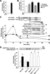
IL-2 modulates glycogen phosphorylase activity in Kit 225 cells through Rac1. A, extracts from unstimulated (−) or stimulated cells (+) (500 units/ml IL-2 for 10 min) were used to measure glycogen phosphorylase activity as described under “Experimental Procedures.” Results show the mean of four independent experiments ±S.D. (**, p < 0.01). B, Kit 225 cells were pretreated with H89 (50 μm) for 1 h and stimulated with forskolin (10 μm; gray columns) or IL-2 (500 units/ml; black columns) for 10 min or left non-stimulated (white column). Cell extracts were used to measure glycogen phosphorylase activity as described under “Experimental Procedures.” Results show the mean of three independent experiments ±S.D. (*, p < 0.05; **, p < 0.01). C, Kit 225 cells were co-transfected with plasmids coding for HA-PYGM-FL, AU5-Rac1 WT, or FLAG-Rac1T17N and HA-PYGMΔRBD with AU5-Rac1 WT. Glycogen phosphorylase activity was measured in non-stimulated cells or cells stimulated with IL-2 for the indicated times. Expression levels of transfected constructs were analyzed by Western blot using antibodies specific for HA, FLAG, and AU5 epitopes as indicated. The results show the mean of three independent experiments ±S.D., and the statistical analysis showed a significant difference (*, p < 0.05). D, Kit 225 cells were transfected with plasmids coding for EGFP-Rac1 WT, EGFP-Rac1T17N, EGFP-Rac1Q61L, EGFP-Rac1F37A+Q61L, or EGFP-Rac1Y40C+Q61L. Glycogen phosphorylase activity was measured in non-stimulated cells or cells stimulated with IL-2 for 10 min. The results show the mean of three independent experiments ±S.D. (*, p < 0.05; **, p < 0.01). As a control, the expression levels of transfected constructs were analyzed by Western blot using GFP-specific antibodies. A, allosteric binding site; CAT, catalytic site; GS, glycogen storage site; RBDGP, Rac1 binding domain of glycogen phosphorylase; S, serine; WB, Western blot.
FIGURE 4.
FIGURE 4.
Rac1/PYGM pathway is necessary for IL-2-dependent Kit 225 proliferation. A, the histogram represents PYGM activity of Kit 225 cells pretreated with 50 μm NCS23766 (Rac1 inhibitor), 10 μm GPI, or vehicle (DMSO) for 16 h and subsequently stimulated with 500 units/ml IL-2 for 10 min at 37 °C. The top panel shows that the Rac1 inhibitor efficiently blocked Rac1 activation in these cells. Pulldown experiments were carried out using GST-RBD of PAK1. Precipitated active Rac1 (Rac1-GTP) and total Rac1 from cell lysates were analyzed by Western blot using anti-Rac1 antibodies. Results show the mean of three independent experiments ± S.D., and the statistical analysis showed a significant difference (**, p < 0.01). B, glycogen accumulation in GPI-treated cells in the presence of IL-2 (16 units/ml). Glycogen was extracted from cells and measured spectrophotometrically using an anthrone-based method as described under “Experimental Procedures.” The glycogen content was corrected for the number of cells present in the sample and is expressed as μg of glycogen/million cells. Results show the mean of three independent experiments ± S.D., and the statistical analysis showed a significant difference (*, p < 0.05). C, cellular proliferation analysis with PKH26 vital dye. Cells stained with PKH26 were cultured in the presence or absence of Rac1 and PYGM inhibitors as described under “Experimental Procedures.” The cell fluorescence level was analyzed on a FACSCalibur cytometer, and data were analyzed using ModFit LT 3.0 software. The curve represents cell number over time (days). Results show the mean of three independent experiments ± S.D. D, proliferation analysis of transfected cells with the vital dye PKH26. Cells transfected with the empty vector pEGFP-C2 or the different gene-expressing vectors, pEGFP-RBD-PAK1, pEGFP-Rac1, pEGFP-PYGM, and pEGFP-PYGMΔRBD, were maintained in the absence of IL-2 for 48 h and stained with PKH26. Fluorescence was analyzed before adding IL-2 (− IL-2 (0 h)) and after 48-h incubation with IL-2 (+ IL-2 (48 h)). Results show the mean of three independent experiments ±S.D., and the statistical analysis showed a significant difference (*, p < 0.05). WB, Western blot.

References

    1. Parri M., Chiarugi P. (2010) Rac and Rho GTPases in cancer cell motility control. Cell Commun. Signal. 8, 23. - PMC - PubMed
    1. Beier I., Düsing R., Vetter H., Schmitz U. (2008) Epidermal growth factor stimulates Rac1 and p21-activated kinase in vascular smooth muscle cells. Atherosclerosis 196, 92–97 - PubMed
    1. Terakado M., Gon Y., Sekiyama A., Takeshita I., Kozu Y., Matsumoto K., Takahashi N., Hashimoto S. (2011) The Rac1/JNK pathway is critical for EGFR-dependent barrier formation in human airway epithelial cells. Am. J. Physiol. Lung Cell. Mol. Physiol. 300, L56–L63 - PubMed
    1. Zhang J. S., Koenig A., Young C., Billadeau D. D. (2011) GRB2 couples RhoU to epidermal growth factor receptor signaling and cell migration. Mol. Biol. Cell 22, 2119–2130 - PMC - PubMed
    1. Bäumer A. T., Ten Freyhaus H., Sauer H., Wartenberg M., Kappert K., Schnabel P., Konkol C., Hescheler J., Vantler M., Rosenkranz S. (2008) Phosphatidylinositol 3-kinase-dependent membrane recruitment of Rac-1 and p47phox is critical for α-platelet-derived growth factor receptor-induced production of reactive oxygen species. J. Biol. Chem. 283, 7864–7876 - PubMed

Publication types

MeSH terms