. 2012 May;107(10):2604-14.
doi: 10.1152/jn.00899.2011. Epub 2012 Feb 15.

Developmental regulation of membrane excitability in rat spinal lamina I projection neurons

Affiliations

Developmental regulation of membrane excitability in rat spinal lamina I projection neurons

Jie Li et al. J Neurophysiol. 2012 May.

Abstract

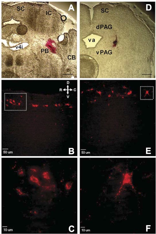
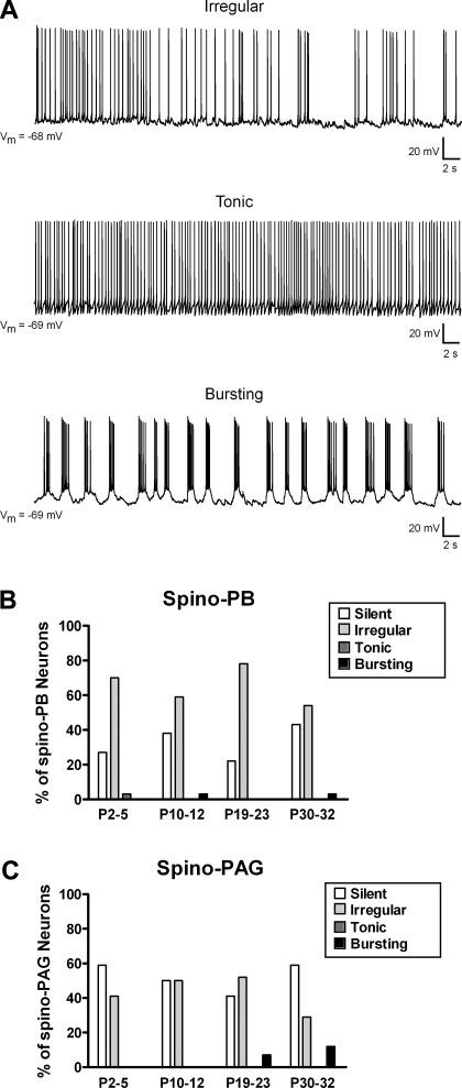
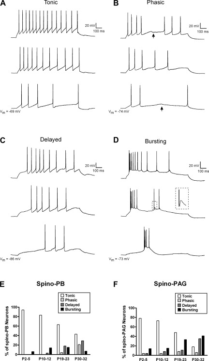
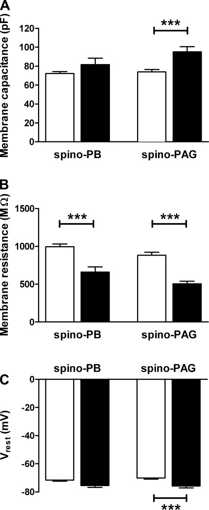
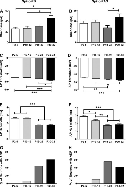
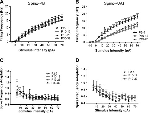
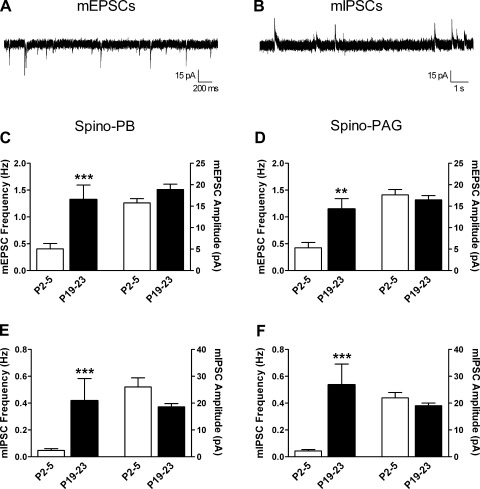
It is now universally recognized that neonates can experience considerable pain. While spinal lamina I neurons projecting to the brain contribute to the generation of hyperalgesia, nothing is known about their electrophysiological properties during early life. Here we have used in vitro whole cell patch-clamp recordings in rat spinal cord slices to determine whether the intrinsic membrane properties of lamina I projection neurons, as well as their synaptic inputs, are developmentally regulated during the early postnatal period. Projection neurons were identified via retrograde transport of DiI injected into the parabrachial nucleus (PB) or periaqueductal gray (PAG) and characterized at postnatal days (P)2-5, P10-12, P19-23, and P30-32. Both spino-PB and spino-PAG neurons demonstrated an age-dependent reduction in spike threshold and duration at room temperature, which was accompanied by a developmental increase in the frequency of miniature excitatory and inhibitory postsynaptic currents. Notably, in both groups, age-dependent changes in the passive membrane properties or rheobase only occurred after the third postnatal week. However, spontaneous activity was significantly more prevalent within the developing spino-PB population and was dominated by an irregular pattern of discharge. In addition, while the instantaneous firing frequency remained unaltered in spino-PB neurons during the first weeks of life, spino-PAG cells fired at a higher rate at P19-23 compared with younger groups, suggesting that the gain of parallel ascending nociceptive pathways may be independently regulated during development. Overall, these results demonstrate that intrinsic membrane excitability is modulated in a cell type-specific manner within developing spinal nociceptive circuits.


Figures

Fig. 1.
Fig. 1.
Retrograde labeling of developing lamina I neurons which project to the parabrachial nucleus (PB) or periaqueductal gray (PAG). A: section of rat brain at postnatal day (P)3 illustrating site where DiI was injected into PB at birth. SC, superior colliculus; IC, inferior colliculus; CB, cerebellum; v4i, fourth ventricle (isthmal). Scale bar, 400 μm. B: sagittal spinal cord section illustrating narrow band of retrogradely labeled neurons within lamina I at P21 after injection of DiI into PB at birth. Orientation arrows indicate dorsal (D), ventral (V), rostral (R), and caudal (C) axes. C: higher magnification of boxed region in B. D: P3 brain section demonstrating location of DiI injection into PAG at birth. dPAG, dorsal PAG; vPAG, ventral PAG; va, aqueduct. Scale bar, 400 μm. E: example of lamina I neurons fluorescently labeled at P21 after injections of DiI into PAG at birth. Same orientation as in B. F: higher magnification of boxed region in E.
Fig. 2.
Fig. 2.
Spontaneous firing patterns in developing lamina I projection neurons. A: spino-PB and spino-PAG neurons located in lamina I of rat spinal cord were classified as irregular (exhibiting intermittent spike activity; top), tonic (continuous firing at a relatively constant frequency; middle), bursting (demonstrating rhythmic burst-firing; bottom), or silent (lack of action potential discharge; not shown). Vm, membrane potential. B and C: patterns of spontaneous activity (SA) in spino-PB (B) and spino-PAG (C) neurons at different postnatal ages, illustrating predominance of irregular spike discharge in both groups throughout development and greater overall prevalence of SA in the spino-PB population. Data for spino-PB group: P2–5: n = 33 cells from 3 rats; P10–12: n = 29 cells from 3 rats; P19–23: n = 27 cells from 4 rats; P30–32: n = 28 cells from 4 rats. Data for spino-PAG group: P2–5: n = 27 cells from 4 rats; P10–12: n = 28 cells from 4 rats; P19–23: n = 27 cells from 5 rats; P30–32: n = 17 cells from 3 rats.
Fig. 3.
Fig. 3.
Evoked action potential (AP) discharge in ascending projection neurons during the early postnatal period. Direct current injection through the patch electrode at increasing intensities (bottom to top) revealed 4 firing patterns in developing spino-PB and spino-PAG lamina I neurons. A: tonic neurons fired APs throughout the 800-ms depolarizations. All traces in panel originate from the same lamina I projection neuron. B: phasic neurons exhibited APs at the beginning of the current step but did not discharge spikes throughout the prolonged depolarization, as irregular gaps in their firing were evident at many stimulus intensities (arrows). C: delayed neurons were distinguished by a long latency to the first spike that varied with stimulus intensity. D: bursting neurons were identified by their slow plateau potentials with superimposed bursts of high-frequency AP discharge. Inset, example of spike afterdepolarization (see boxed region). E and F: when APs were evoked by intracellular current injection from the resting membrane potential, the majority of both spino-PB (E) and spino-PAG (F) neurons exhibited tonic firing during the first 3 postnatal weeks, while a more even distribution of firing patterns was evident by P30–32. Sample sizes were the same as described in Fig. 2.
Fig. 4.
Fig. 4.
Distinct passive membrane properties of ascending projection neurons that demonstrate evoked burst-firing. A: average membrane capacitance of bursting (filled bars) spino-PAG neurons was higher compared with nonbursting cells (open bars) in this population (***P = 0.0002; Mann-Whitney test; right), while no significant differences were noted in the spino-PB group (P = 0.141; left). B: bursting neurons (filled bars) exhibited significantly lower membrane resistance compared with other projection neurons (open bars) in both the spino-PB (***P = 0.0008; Mann-Whitney test; left) and spino-PAG (***P < 0.0001; right) populations. C: resting membrane potential (Vrest) was more hyperpolarized in spino-PAG neurons showing burst-firing in response to current injection compared with spino-PAG cells showing other patterns of evoked discharge (***P = 0.0009; right). Data on bursting neurons originate from 5 rats for the spino-PB group and 12 rats for the spino-PAG population.
Fig. 5.
Fig. 5.
Age-dependent modulation of membrane excitability in developing spinal projection neurons. A and B: minimum current needed to evoke an AP (i.e., rheobase) did not change significantly during early postnatal development in either the spino-PB (A) or spino-PAG (B) population of lamina I neurons, although higher rheobase levels were seen in the P30–32 group (*P < 0.05; Kruskal-Wallis test with Dunn's multiple comparison test). C and D: however, an age-related reduction in AP threshold was observed in both spino-PB (C; *P < 0.05, ***P < 0.001; Kruskal-Wallis test with Dunn's posttest) and spino-PAG (D; *P < 0.05, **P < 0.01, ***P < 0.001; 1-way ANOVA with Tukey's multiple comparison test) neurons. E and F: both groups of projection neurons exhibited a developmental decrease in spike duration (*P < 0.05, **P < 0.01, ***P < 0.001; Kruskal-Wallis test with Dunn's posttest). G and H: spino-PB (G) and spino-PAG (H) cells also demonstrated an increased prevalence of spike afterdepolarizations (ADPs) with age (P < 0.0001; χ2-test). Sample sizes were the same as described in Fig. 2.
Fig. 6.
Fig. 6.
Firing rate of spino-PAG, but not spino-PB, projection neurons accelerates during postnatal development. A: plot of mean instantaneous firing frequency as a function of stimulus intensity in spino-PB neurons reveals no significant differences in repetitive firing between age groups (P > 0.05; 2-way ANOVA; n = 12–30 in each group). B: in contrast, P19–23 spino-PAG neurons fired at a significantly higher frequency compared with younger ages (#P < 0.05, ##P < 0.01 vs. P10–12; &P < 0.05 vs. P2–5; **P < 0.01, ***P < 0.001 vs. both ages; 2-way ANOVA with Bonferroni posttests; n = 14–20 in each group). Spino-PAG neurons were not analyzed at P30–32 because of the low number (n = 3) of tonically firing neurons observed at this age. C and D: the degree of spike frequency adaptation across a range of stimulus intensities did not change significantly with age in either population of lamina I projection neurons (P > 0.05; 2-way ANOVA). The numbers of animals used were the same as described in Fig. 2.
Fig. 7.
Fig. 7.
Developmental increase in the efficacy of spontaneous excitatory and inhibitory synaptic transmission onto spinal projection neurons. A: example of miniature excitatory postsynaptic currents (mEPSCs) isolated at a holding potential of −70 mV in an immature lamina I projection neuron identified by retrograde transport of DiI. B: example of miniature inhibitory postsynaptic currents (mIPSCs) recorded in the same neuron from a holding potential of 0 mV. C and D: frequency of mEPSCs increased with age in both spino-PB (***P = 0.0003; Mann-Whitney test; C, left) and spino-PAG (**P = 0.0013; D, left) neurons without a significant change in mEPSC amplitude (right). E and F: similarly, mIPSC frequency was significantly higher at P19–23 compared with P2–5 in the same populations of spino-PB (***P < 0.0001; Mann-Whitney; E, left) and spino-PAG neurons (***P = 0.0002; F, left), while mIPSC amplitude was unaltered (right). Data for spino-PB group are derived from 3 rats at P2–5 and 4 rats at P19–23. Data for spino-PAG group are derived from 3 rats at P2–5 and 4 rats at P19–23.

Similar articles

Cited by

References

    1. Altman J, Bayer SA. Atlas of Prenatal Rat Brain Development. Boca Raton, FL: CRC, 1995
    1. Andersen P, Sundberg SH, Sveen O, Swann JW, Wigstrom H. Possible mechanisms for long-lasting potentiation of synaptic transmission in hippocampal slices from guinea-pigs. J Physiol 302: 463–482, 1980 - PMC - PubMed
    1. Andrew D. Quantitative characterization of low-threshold mechanoreceptor inputs to lamina I spinoparabrachial neurons in the rat. J Physiol 588: 117–124, 2010 - PMC - PubMed
    1. Baccei ML, Bardoni R, Fitzgerald M. Development of nociceptive synaptic inputs to the neonatal rat dorsal horn: glutamate release by capsaicin and menthol. J Physiol 549: 231–242, 2003 - PMC - PubMed
    1. Baccei ML, Fitzgerald M. Development of GABAergic and glycinergic transmission in the neonatal rat dorsal horn. J Neurosci 24: 4749–4757, 2004 - PMC - PubMed

Publication types