. 2012 Mar 6;51(9):1996-2007.
doi: 10.1021/bi201799a. Epub 2012 Feb 27.

Kinetics and fidelity of polymerization by DNA polymerase III from Sulfolobus solfataricus

Affiliations

Kinetics and fidelity of polymerization by DNA polymerase III from Sulfolobus solfataricus

Robert J Bauer et al. Biochemistry. .

Abstract

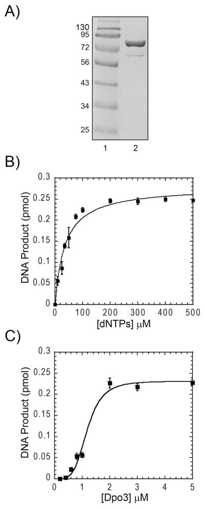
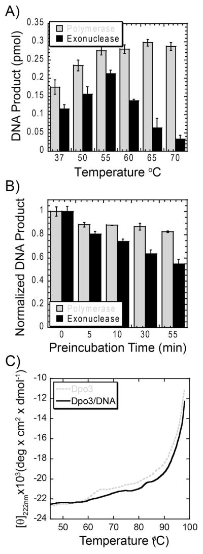
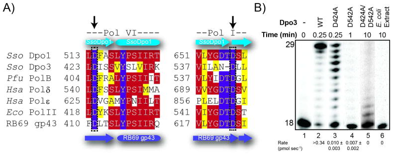
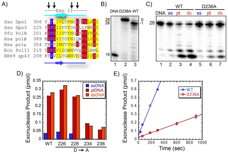
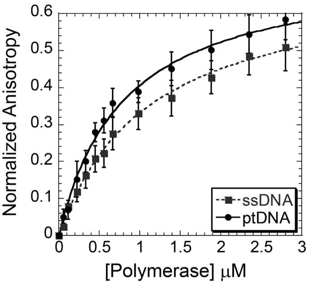
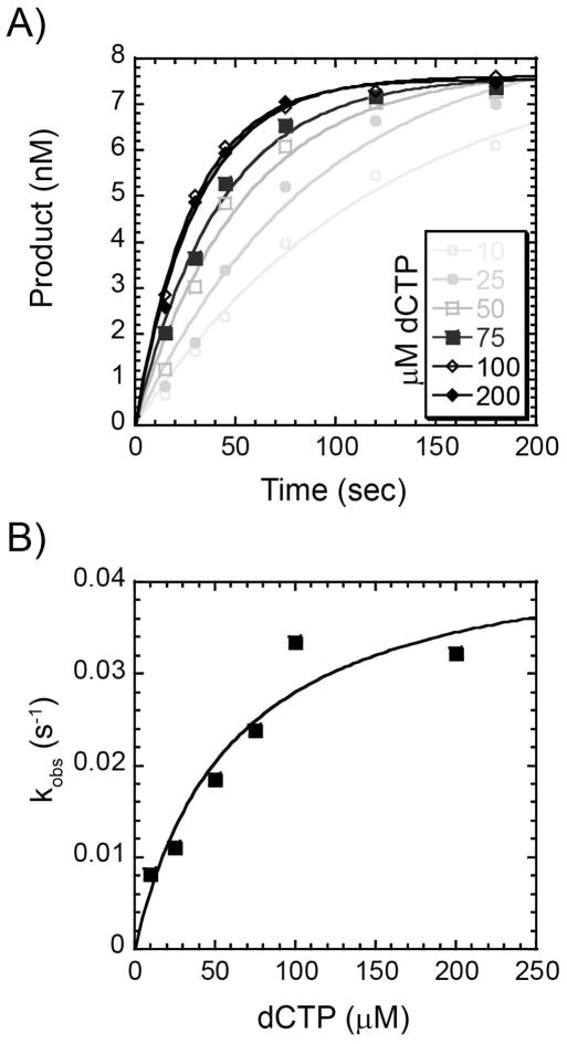
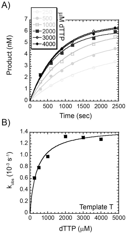
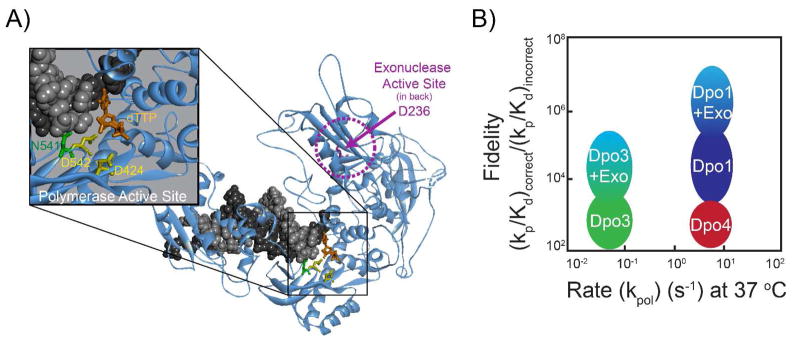
We have biochemically and kinetically characterized the polymerase and exonuclease activities of the third B-family polymerase (Dpo3) from the hyperthermophilic Crenarchaeon, Sulfolobus solfataricus (Sso). We have established through mutagenesis that despite incomplete sequence conservation, the polymerase and exonuclease active sites are functionally conserved in Dpo3. Using pre-steady-state kinetics, we can measure the fidelity of nucleotide incorporation by Dpo3 from the polymerase active site alone to be 10(3)-10(4) at 37 °C. The functional exonuclease proofreading active site will increase fidelity by at least 10(2), making Dpo3 comparable to other DNA polymerases in this family. Additionally, Dpo3's exonuclease activity is modulated by temperature, where a loss of promiscuous degradation activity can be attributed to a reorganization of the exonuclease domain when it is bound to primer-template DNA at high temperatures. Unexpectedly, the DNA binding affinity is weak compared with those of other DNA polymerases of this family. A comparison of the fidelity, polymerization kinetics, and associated functional exonuclease domain with those previously reported for other Sso polymerases (Dpo1 and Dpo4) illustrates that Dpo3 is a potential player in the proper maintenance of the archaeal genome.


Figures

Figure 1
Figure 1
(A) Purified Dpo3 on a Coomassie-stained SDS-PAGE gel: lane 1, protein markers, and lane 2, Dpo3 (88 kDa). (B) Optimization of dNTP concentrations on total DNA product produced by 2 μM Dpo3 on ptDNA in Tris (pH = 7.5), 10 mM Mg2+ at 70 °C. The Km for dNTPs (36 ± 5 μM) was determined from the average of three independent experiments fit to Equation 1. (C) Effect of Dpo3 concentration on total DNA product produced using long ptDNA template. Reactions were performed in Tris (pH = 7.5), 200 μM dNTPs, 10 mM Mg2+ at 70 °C for 10 minutes. The apparent dissociation constant (K’d) for Dpo3 activity (1.2 ± 0.1 μM) was determined from the average of three independent experiments fit to Equation 1. The cooperativitiy parameter (n) was equal to 4.4 ± 1.1.
Figure 2
Figure 2
(A) Temperature dependence of 2 μM Dpo3 (WT) in 50 mM Tris (pH = 7.5), 10 mM Mg2+, 200 μM dNTPs on polymerase (grey) and exonuclease (black) activities on ptDNA (36 nM) in a 10 minute reaction. The reported values and errors are the average of three independent experiments. (B) Thermostability of 2μM Dpo3 (WT) after preincubation at 70 °C for the indicated time points. Quantification of product formation for polymerase activity (grey) after the addition of dNTPs and exonuclease activity (black) after the addition of ptDNA, in a 10 minute reaction at 70 °C (polymerase activity) or 55 °C (exonuclease activity). The error bars represent the standard error from at least three independent experiments. (C) Thermal melting of Dpo3 alone (grey, dashed) or bound (black, solid) to hairpin DNA monitored by circular dichroism at 222 nm (2 °C increments).
Figure 3
Figure 3
(A) Amino acid alignment of DNA polymerase domains VI and I using CLUSTAL W2 (http://www.ebi.ac.uk/Tools/clustalw2) for common members within the B-family of DNA replication polymerases. Color coding for slightly (yellow), mostly (red) and absolutely (purple) conserved residues are indicated. The secondary structure elements are derived from the crystal structure of Dpo1 (cyan) (PDBID: 1S5J) or RB69 gp43 (purple) (PDBID: 1CLQ). Species are each listed as a three letter code: Sso, Sulfolobus solfataricus, Pfu, Pyrococcus furiosus, Hsa, Homo sapien, and Eco, Escherichia coli. Arrows represent the residues in Dpo3 that were mutated and constitute the active site aspartates. (B) Effect of single (D424A or D542A) or double mutants (D424A/D542A) of Dpo3 on the extension of Template (T)G at 60 °C for indicated times in optimal buffer conditions. The average rates in pmol sec−1 from multiple independent experiments are listed below the corresponding lanes of the gel.
Figure 4
Figure 4
(A) Amino acid alignment of exonuclease domain II using CLUSTAL W2 (http://www.ebi.ac.uk/Tools/clustalw2) for common members within the B-family of DNA replication polymerases. Color coding for slightly (yellow), mostly (red) and absolutely (purple) conserved residues are indicated. The secondary structure elements are derived from the crystal structure of Dpo1 (cyan) (PDBID: 1S5J) or RB69 gp43 (purple) (PDBID: 1CLQ). Species are each listed as a three letter code: Sso, Sulfolobus solfataricus, Pfu, Pyrococcus furiosus, Hsa, Homo sapien and Eco, Escherichia coli. Arrows represent the residues in Dpo3 that were mutated. (B) Polymerase reactions comparing full length product formation from WT and D236A Dpo3 on Template T at 70 °C for three minutes, showing major products (29 base, blunt, 0) or (28 base, recessed, −1) for D236A or WT Dpo3, respectively. C) Exonuclease experiment on single strand (ssDNA), primer template (ptDNA), and double-strand (dsDNA) DNA for both WT and D236A Dpo3 at 55 °C for 10 minutes. (D) Quantification of exonuclease cleavage products for WT Dpo3 and each prospective exonuclease mutant (D226A, D228A, D234A, D236A), on all three DNA conformations: ssDNA (blue), ptDNA (red), or dsDNA (orange) at 55 °C for 10 minutes. (E) Quantification of the steady-state rate of exonuclease products produced from ptDNA by WT (0.031 ± 0.001 pmol s−1) or D236A (0.0046 ± 0.0003 pmol s−1) Dpo3 from at least two independent experiments. The error bars represent the standard error of the reaction.
Figure 5
Figure 5
Change in fluorescence anisotropy upon Dpo3 (D236A) binding to either Cy3-labeled ssDNA (Cy3DNA) (grey, dash) or ptDNA (black, solid). Data points were fit to Equation 4 to extract a Kd of binding for ssDNA (1.10 ± 0.08 μM) and ptDNA (0.81 ± 0.06 μM). The error bars represent the standard error from at least three independent experiments.
Figure 6
Figure 6
Concentration dependence of the pre-steady state rate of correct nucleotide incorporation. (A) A preincubated solution containing 2 μM Dpo3 and Template G (9.6 nM) was mixed with increasing concentrations of dCTP (10 μM – 200 μM) for the indicated time points. The data was fit to Equation 3 to determine the single-exponential rate (kobs). (B) kobs values were plotted as a function of dCTP concentration and fit with Equation 4 to yield kp (0.045 ± 0.008 s−1) and Kd (61 ± 26 μM).
Figure 7
Figure 7
Concentration dependence of the pre-steady state rate of incorrect nucleotide incorporation. (A) A preincubated solution containing 2 μM Dpo3 and Template T (9.6 nM) was mixed with increasing concentrations of dTTP (250 μM – 4 mM) for the indicated time points. The data was fit to Equation 3 to determine the single exponential rate (kobs). (B) kobs values were plotted as a function of dTTP concentration and fit with Equation 4 to yield kp (0.0015 ± 0.0001 s−1) and Kd (0.39 ± 0.08 mM).
Figure 8
Figure 8
A) Homology model of the ternary complex of Dpo3 bound to DNA (black/grey) with incoming dTTP (orange) highlighting the aspartates in the polymerase (D424 and D542, yellow) and exonuclease (D236, pink) active sites. N541 (green) is also shown orientated towards the minor groove of the dsDNA template. B) Graphical representation of the fidelity [(kp/Kd)correct/(kp/Kd)incorrect] as a function of the rate (kpol) comparing Dpo1, Dpo3, and Dpo4.

Similar articles

Cited by

References

    1. Filee J, Forterre P, Sen-Lin T, Laurent J. Evolution of DNA polymerase families: evidences for multiple gene exchange between cellular and viral proteins. J Mol Evol. 2002;54:763–773. - PubMed
    1. Burgers PM, Koonin EV, Bruford E, Blanco L, Burtis KC, Christman MF, Copeland WC, Friedberg EC, Hanaoka F, Hinkle DC, Lawrence CW, Nakanishi M, Ohmori H, Prakash L, Prakash S, Reynaud CA, Sugino A, Todo T, Wang Z, Weill JC, Woodgate R. Eukaryotic DNA polymerases: Proposal for a revised nomenclature. J Biol Chem. 2001;276:43487–43490. - PubMed
    1. Hubscher U, Maga G, Spadari S. Eukaryotic DNA polymerases. Annu Rev Biochem. 2002;71:133–163. - PubMed
    1. Bebenek K, Kunkel TA. Functions of DNA polymerases. Adv Protein Chem. 2004;69:137–165. - PubMed
    1. Kunkel TA, Bebenek K. DNA replication fidelity. Annu Rev Biochem. 2000;69:497–529. - PubMed

Publication types