Review
. 2012 Apr 5;4(3):e00082.
doi: 10.1042/AN20120010.

Neurological diseases as primary gliopathies: a reassessment of neurocentrism

Affiliations
Review

Neurological diseases as primary gliopathies: a reassessment of neurocentrism

Alexei Verkhratsky et al. ASN Neuro. .

Abstract

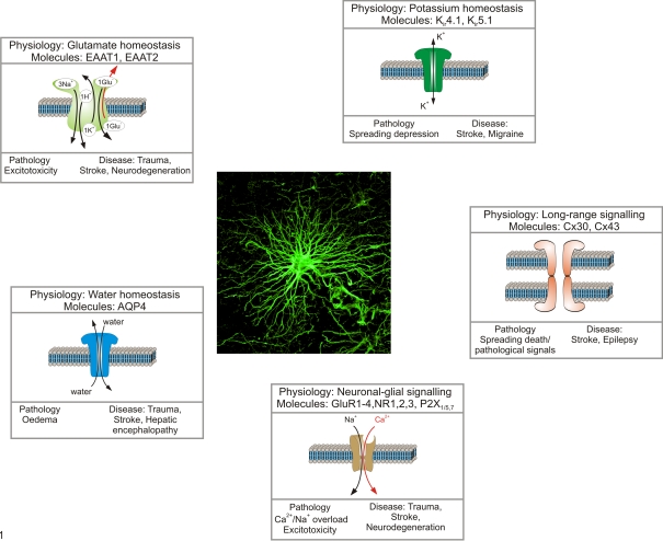
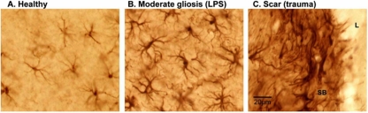
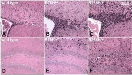
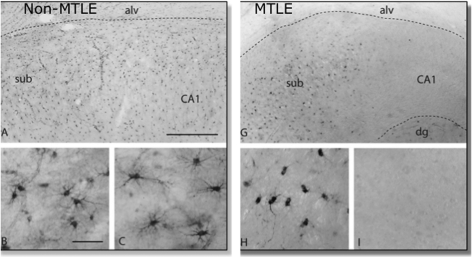
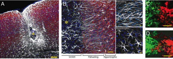
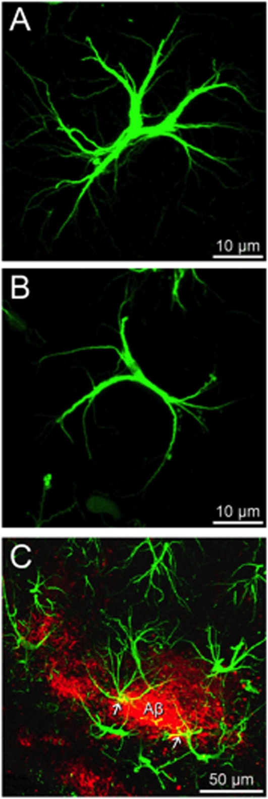
Diseases of the human brain are almost universally attributed to malfunction or loss of nerve cells. However, a considerable amount of work has, during the last decade, expanded our view on the role of astrocytes in CNS (central nervous system), and this analysis suggests that astrocytes contribute to both initiation and propagation of many (if not all) neurological diseases. Astrocytes provide metabolic and trophic support to neurons and oligodendrocytes. Here, we shall endeavour a broad overviewing of the progress in the field and forward the idea that loss of homoeostatic astroglial function leads to an acute loss of neurons in the setting of acute insults such as ischaemia, whereas more subtle dysfunction of astrocytes over periods of months to years contributes to epilepsy and to progressive loss of neurons in neurodegenerative diseases. The majority of therapeutic drugs currently in clinical use target neuronal receptors, channels or transporters. Future therapeutic efforts may benefit by a stronger focus on the supportive homoeostatic functions of astrocytes.


Figures

Figure 1
Figure 1. General pathophysiology of astroglia
The homoeostatic cascades expressed in astrocytes control various aspects of CNS homoeostasis including extracellular ion homoeostasis (K+ buffering via Kir channels, Na/K pump and K transporters), regulate movements and distribution of water (via aquaporins and connexins), control extracellular concentration of neurotransmitters (by dedicated transporters) and provide the main reactive oxygen species scavenging system. In pathological conditions, the very same systems may contribute to brain damage. Failure in water transport triggers brain oedema, reversal of neurotransmitter transporters contributes to glutamate excitotoxicity, inadequate K+ buffering promotes further overexcitation of neural cells and spreading depression, and connexins become a conduit for death signals.
Figure 2
Figure 2. Appearance of astrocytes and different types of reactive astrocytes in mouse cerebral cortex
Images show immunohistochemistry for the intermediate filament protein, GFAP, which visualizes the cell cytoskeleton. (A) In healthy cortex, some, but not all astrocytes express detectable levels of GFAP; cell bodies are small and the cytoskeleton is thin and restricted largely to the proximal portions of cell processes. (B) In response to the bacterial antigen, lipopolysaccharide (LPS) injected into the lateral cerebral ventricle, cortical astrocytes become moderately reactive, with up-regulation of GFAP expression such that it is now detectable in all astrocytes. In addition, there is substantial hypertrophy of the astrocyte cell bodies as well as hypertrophy of stem processes and associated cytoskeleton. However, there is no astrocyte proliferation and individual cells continue to respect their individual, non-overlapping domains. (C) In response to a severe traumatic injury that creates a lesion (L) with tissue necrosis and invasion of inflammatory cells, astrocytes not only become reactive but also proliferate in the immediate vicinity of the lesion and form a scar with a dense scar border (SB) that comprises many newly generated astrocytes that do not exhibit individual domains and instead have many overlapping and intermingling processes. All images are at the same magnification. Scale bar = 20 μm (Photos courtesy of the Sofroniew laboratory).
Figure 3
Figure 3. Immunostaining for GFAP in brain tissue from mouse models of Alexander disease showing abundant Rosenthal fibres in the periventricular region
GFAP immunohistochemistry in the periventricular white matter of (A) wild-type or (B, C) knock-in point mutants expressing either R76H or R236H mutant forms of GFAP (equivalent to the common R79H and R239H mutations in human GFAP). Abundant Rosenthal fibres with increased immunoreactivity for GFAP are particularly evident in periventricular and white matter astrocytes of adult mice (3 months old). Reproduced with permission from Figures 3(A)–3(C) of Hagemann et al. (2006) ©2006 Society for Neuroscience.
Figure 4
Figure 4. Expression of glutamine synthetase immunoreactivity in a non-sclerotic and sclerotic hippocampus
Glutamine synthetase immunoreactivty in the subiculum/CA1 region in a non-sclerotic [non-MTLE (mesial temporal lobe epilepsy)] (A) and sclerotic (MTLE) (G) hippocampus. Neurologically normal autopsy hippocampus shows a pattern of staining exactly similar to (A). (B, C) in higher magnification shows GS immunopositive astrocytes in both the subiculum and CA1 area of a normal or non-sclerotic hippocampus. The sclerotic hippocampus, in which the subiculum does not have neuronal loss shows GS immunoreactive astrocytes (H), whereas the neuron-depleted astrocyte-rich CA1 area (I) shows depletion of GS in astrocytes. (A and G) Scale bar = 0.5 mm. (B, C, H and I) Scale bar = 100 μm. Reprinted from The Lancet, 363, Eid T, Thomas MJ, Spencer DD, Runden-Pran E, Lai JC, Malthankar GV, Kim JH, Danbolt NC, Ottersren OP, de Lanerolle NC, Loss of glutamine synthetase in the human epileptogenic hippocampus: possible mechanism for raised extracellular glutamate in mesial temporal lobe epilepsy, 28-37, Copyright (2004), with permission from Elsevier.
Figure 5
Figure 5. Organization of reactive astrocytes in a model of post-traumatic epilepsy induced by cortical injection of a ferrous chloride solution
(A) Site of cortical injury 6 months after injury. The centre of the lesion (yellow asterisk) is surrounded by palisading astrocytes and, at a greater distance, by hypertrophic astrocytes. The mouse exhibited daily multiple generalized grand mall seizures. (B) Higher power image of a similar lesion displaying palisading and hyperthrophic astrocytes. White, GFAP; red, Map2; blue, Sytox. (C, D) Neighbouring astrocytes in control, non-EL brain exhibit little overlap of processes (C), whereas extensive overlap of processes between two adjacent astrocytes is evident in a mouse with epilepsy (D). Neighbouring astrocytes were duolistically labelled with DiL (green) or DiD (ref). Scale bar = 100 μm (A), 50 μm and 10 μm (B), 10 μm (C, D). See Oberheim et al. (2008) for details.
Figure 6
Figure 6. Astrocytes in neurodegeneration
(A) Fluorescence micrographs illustrating a normal hippocampal astrocyte labelled with anti-GFAP antibody with elongated and multiple radial processes in an old (18 months) control animal. (B) In age-matched 3×TG-AD animals, astrocytes show a morphological atrophy with a significant reduction in cell soma volume and area as well as a reduction in the number and width of processes. (C) Confocal image showing hypertrophic astrocytes (green) concentrated around Aβ plaques (red); occasionally some of the astrocytes show intracellular Aβ accumulation (yellow). Scale bar (A and B): 10 μm; (C): 50 μm.

Similar articles

Cited by

References

    1. Abbott NJ. Dynamics of CNS barriers: evolution, differentiation, and modulation. Cell Mol Neurobiol. 2005;25:5–23. - PMC - PubMed
    1. Alexander WS. Progressive fibrinoid degeneration of fibrillary astrocytes associated with mental retardation in a hydrocephalic infant. Brain. 1949;72:373–381. - PubMed
    1. Allaman I, Gavillet M, Belanger M, Laroche T, Viertl D, Lashuel HA, Magistretti PJ. Amyloid-β aggregates cause alterations of astrocytic metabolic phenotype: impact on neuronal viability. J Neurosci. 2010;30:3326–3338. - PMC - PubMed
    1. Alvarez-Buylla A, Garcia-Verdugo JM, Tramontin AD. A unified hypothesis on the lineage of neural stem cells. Nat Rev Neurosci. 2001;2:287–293. - PubMed
    1. Alzheimer A, Nissl F, Alzheimer A. Beiträge zur Kenntnis der pathologischen Neuroglia und ihrer Beziehungen zu den Abbauvorgängen im Nervengewebe. Histologische und Histopathologische Arbeiten über die Grosshirnrinde mit besonderer Berücksichtigung der pathologischen Anatomie der Geisteskrankheiten. Verlag von Gustav Fischer Jena. 1910:401–562.

Publication types

MeSH terms