Review
. 2012 May;36(3):734-47.
doi: 10.1111/j.1574-6976.2012.00333.x. Epub 2012 Mar 13.

Host cell invasion by Trypanosoma cruzi: a unique strategy that promotes persistence

Affiliations
Review

Host cell invasion by Trypanosoma cruzi: a unique strategy that promotes persistence

Maria Cecilia Fernandes et al. FEMS Microbiol Rev. 2012 May.

Abstract

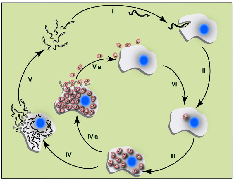
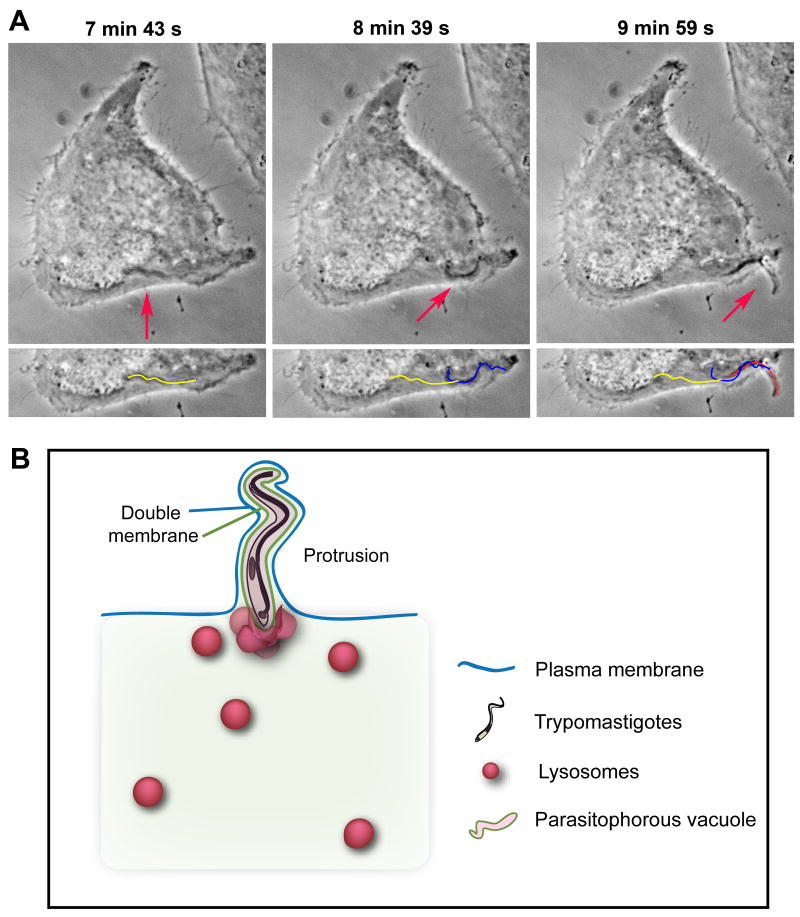
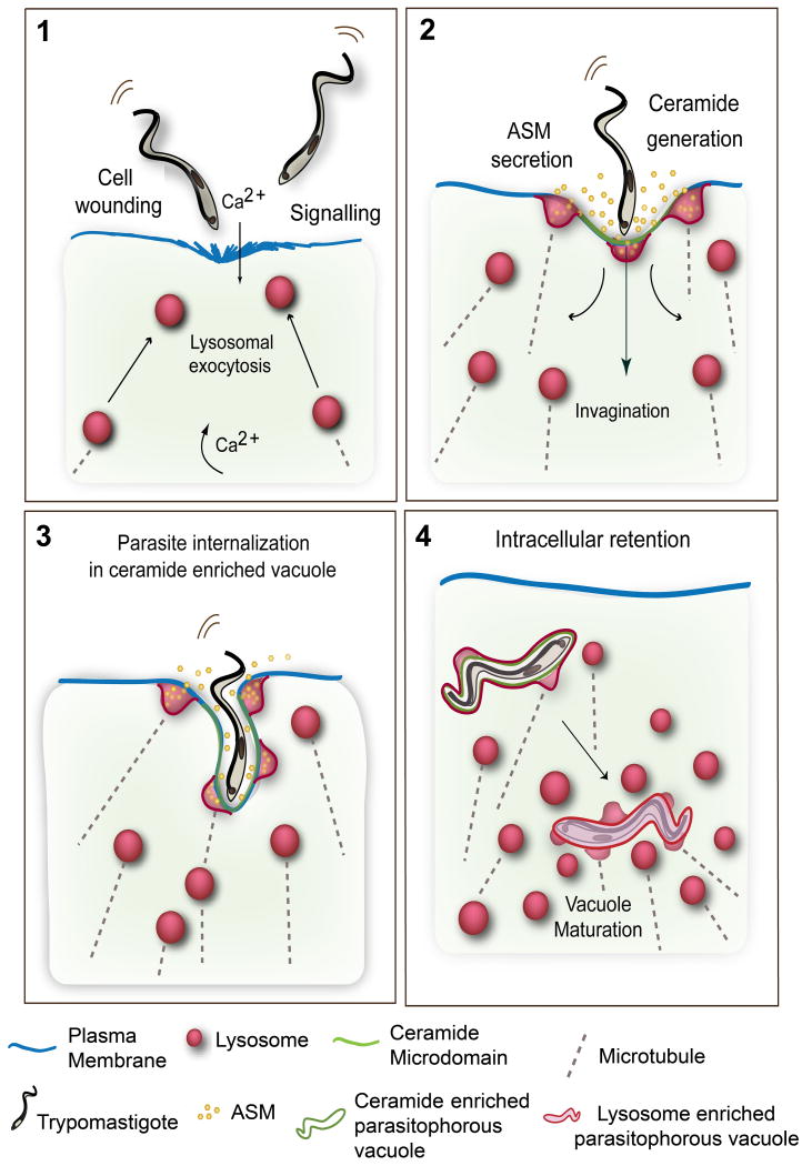
The intracellular protozoan parasite Trypanosoma cruzi is the causative agent of Chagas' disease, a serious disorder that affects millions of people in Latin America. Despite the development of lifelong immunity following infections, the immune system fails to completely clear the parasites, which persist for decades within host tissues. Cardiomyopathy is one of the most serious clinical manifestations of the disease, and a major cause of sudden death in endemic areas. Despite decades of study, there is still debate about the apparent preferential tropism of the parasites for cardiac muscle, and its role in the pathology of the disease. In this review, we discuss these issues in light of recent observations, which indicate that T. cruzi invades host cells by subverting a highly conserved cellular pathway for the repair of plasma membrane lesions. Plasma membrane injury and repair is particularly prevalent in muscle cells, suggesting that the mechanism used by the parasites for cell invasion may be a primary determinant of tissue tropism, intracellular persistence, and Chagas' disease pathology.


Figures

Figure 1
Figure 1. Trypanosoma cruzi replication in the mammalian host
Flagellated infective forms of the parasite (trypomastigotes) invade mammalian host cells (I) and differentiate into round intracellular parasites (amastigotes) (II), which replicate by binary fission in the cytoplasm (III). Amastigotes can then follow two distinct paths: 1) Parasites differentiate into trypomastigotes, which disrupt the host cell (IV) and invade neighboring cells or enter the bloodstream, disseminating the infection (V); 2) Heavily infected cells may be disrupted prematurely (particularly once immunity is established) releasing amastigotes that can re-invade cells by phagocytosis, thus contributing to parasite persistence (IVa, Va and VI).
Figure 2
Figure 2. Internalized trypomastigotes protrude from host cells
(A) Time Lapse live imaging (phase-contrast) of a trypomastigote moving inside a HeLa cell, and forming a plasma membrane protrusion (arrows). The yellow line represents the parasite's position at 7 min 43 s, the blue line at 8 min 39 s, and red line at 9 min 59 s (bottom panel). (B) Schematic model for protrusion event. Protruding trypomastigotes are surrounded by two distinct membranes: parasitophorous vacuole (green line), and plasma membrane (blue line). The parasites protrude in the direction of their flagellar movement (anterior end pointing outward) and stretch the overlying plasma membrane, while lysosomes progressively fuse with the vacuole and ultimately anchor trypomastigotes inside the cell.
Figure 3
Figure 3. Parasitophorous Vacuoles of recently internalized trypomastigotes are enriched in ceramide
(A) Confocal image of an infected HeLa cell (15 min of infection) incubated with anti-ceramide (red) and anti-Lamp1 (green) antibodies. A protruding trypomastigote outlined with ceramide patches is seen on the left, while Lamp-1 enriched lysosomes – the source of acid sphingomyelinase - are observed closely associated with the parasitophorous vacuole (arrows). The DAPI-stained parasite kinetoplast (arrowhead) and the host cell and parasite nuclei are shown in blue. (B) 3D reconstruction of (A) highlighting the ceramide-enriched microdomains and the lysosomes.
Figure 4
Figure 4. Model for T. cruzi cell invasion mediated by wounding and plasma membrane repair
(1) Trypomastigotes wound mammalian cells, causing influx of extracellular Ca2+ and exocytosis of lysosomes. Signaling events also generate cytosolic Ca2+ transients. (2) Extracellular release of lysosomal ASM generates ceramide on the outer leaflet of the plasma membrane. (3) Ceramide-enriched plasma membrane microdomains facilitate trypomastigote internalization, while lysosomes continue to fuse with the nascent parasitophorous vacuoles, delivering ASM and generating additional ceramide. Lysosomal membrane anchoring to microtubules provides the force for pulling the parasites into the cells. (4) Recently internalized trypomastigotes residing in ceramide/Lamp1-enriched parasitophorous vacuoles fuse with additional lysosomes leading to parasite retention inside the cell.

References

    1. Alves MJ, Colli W. Trypanosoma cruzi: adhesion to the host cell and intracellular survival. IUBMB Life. 2007;59:274–279. - PubMed
    1. Alves MJ, Mortara RA. A century of research: what have we learned about the interaction of Trypanosoma cruzi with host cells? Mem I Oswaldo Cruz. 2009;104 1:76–88. - PubMed
    1. Andrade LO, Andrews NW. Lysosomal fusion is essential for the retention of Trypanosoma cruzi inside host cells. J Exp Med. 2004;200:1135–1143. - PMC - PubMed
    1. Andrade LO, Andrews NW. The Trypanosoma cruzi-host-cell interplay: location, invasion, retention. Nat Rev Microbiol. 2005;3:819–823. - PubMed
    1. Andrews NW, Whitlow MB. Secretion by Trypanosoma cruzi of a hemolysin active at low pH. Mol Biochem Parasitol. 1989;33:249–256. - PubMed

Publication types