Comparative Study
. 2012 Feb 21;59(8):751-63.
doi: 10.1016/j.jacc.2011.10.888.

Catheter-deliverable hydrogel derived from decellularized ventricular extracellular matrix increases endogenous cardiomyocytes and preserves cardiac function post-myocardial infarction

Affiliations
Comparative Study

Catheter-deliverable hydrogel derived from decellularized ventricular extracellular matrix increases endogenous cardiomyocytes and preserves cardiac function post-myocardial infarction

Jennifer M Singelyn et al. J Am Coll Cardiol. .

Abstract

Objectives: This study evaluated the use of an injectable hydrogel derived from ventricular extracellular matrix (ECM) for treating myocardial infarction (MI) and its ability to be delivered percutaneously.

Background: Injectable materials offer promising alternatives to treat MI. Although most of the examined materials have shown preserved or improved cardiac function in small animal models, none have been specifically designed for the heart, and few have translated to catheter delivery in large animal models.

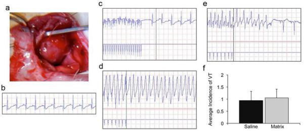
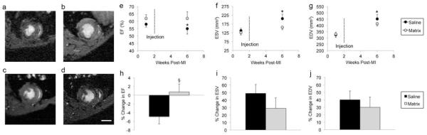
Methods: We have developed a myocardial-specific hydrogel, derived from decellularized ventricular ECM, which self-assembles when injected in vivo. Female Sprague-Dawley rats underwent ischemia reperfusion followed by injection of the hydrogel or saline 2 weeks later. The implantation response was assessed via histology and immunohistochemistry, and the potential for arrhythmogenesis was examined using programmed electrical stimulation 1 week post-injection. Cardiac function was analyzed with magnetic resonance imaging 1 week pre-injection and 4 weeks post-MI. In a porcine model, we delivered the hydrogel using the NOGA-guided MyoStar catheter (Biologics Delivery Systems, Irwindale, California), and utilized histology to assess retention of the material.

Results: We demonstrate that injection of the material in the rat MI model increases endogenous cardiomyocytes in the infarct area and maintains cardiac function without inducing arrhythmias. Furthermore, we demonstrate feasibility of transendocardial catheter injection in a porcine model.

Conclusions: To our knowledge, this is the first in situ gelling material to be delivered via transendocardial injection in a large animal model, a critical step towards the translation of injectable materials for treating MI in humans. Our results warrant further study of this material in a large animal model of MI and suggest this may be a promising new therapy for treating MI.


Figures

Figure 1
Figure 1. Myocardial matrix fabrication
(a) Porcine ventricular myocardium is sliced, and then (b) decellularized using SDS. (c) H&E staining of a histological section reveals cellular removal. The decellularized ECM is then milled into a fine powder (d), and solubilized through enzymatic digestion (e), which allows for injection via syringe and a 27G needle (f).
Figure 2
Figure 2. Programmed electrical stimulation to assess potential for arrhythmogenesis
(a) An electrode paces the LV of rat hearts one week post-injection. (b) Intrinsic rhythm of rat heart prior to pacing. (c) Return to intrinsic rhythm when burst pacing is stopped. (d) Sustained VT following a single extra-stimulus. (e) Non-sustained VT following a single extra-stimulus. (f) There was no observed difference in the average incidence of VT between saline and myocardial matrix groups, demonstrating that the myocardial matrix hydrogel is not pro-arrhythmogenic.
Figure 3
Figure 3. Histological and immunohistochemical assessment
(a) H&E of matrix injected infarct demonstrating moderate mononuclear cell infiltration (scale bar = 50 μm). (b) An increased average area of cardiomyocyte islands were found in the infarct of myocardial matrix injected hearts compared to saline controls (*P<0.05). An island of viable myocardium in the infarct of a matrix injected heart is denoted by the arrow (scale bar = 200 μm). Cardiac Troponin T is shown in green with the infarct and borderzone denoted by I and BZ, respectively. (c) There was no significant difference in the number of infiltrating M2 macrophages between groups. Nuclei are stained blue with Hoechst and CD163+ cells are labeled red with a few cells denoted by the arrow points (scale bar = 100 μm). (d) An increase in proliferative cells were observed in matrix injected hearts (*P<0.05). Nuclei are stained blue with Hoechst and Ki67+ cells are labeled red. Co-stained nuclei appear purple with a couple cells denoted by arrow points (scale bar = 50 μm). (e) C-kit+ cells were observed in low numbers within the myocardial matrix scaffold. C-kit+ cells (denoted with arrow points) are labeled in green, with nuclei in blue (scale bar = 10 μm).
Figure 4
Figure 4. MRI analysis in rat myocardial infarction model
(a) Baseline (1 week post-MI, 1 week pre-injection) and (b) 4 weeks post-injection images of saline injected heart. (c) Baseline and (d) 4 weeks post-injection images of myocardial matrix injected heart. Scale bar = 0.5 cm. (e) Ejection fraction of saline controls significantly declined (p = 0.04), while the (f) end-systolic volume (p = 0.01) and (g) end-diastolic volume (p = 0.01) significantly expanded between 1 week and 6 weeks post-MI. In contrast, there was no statistical significance for EF (p = 0.8), ESV (p = 0.054), and EDV (p = 0.06) between time points in the myocardial matrix group. Myocardial matrix injected animals had an overall increase in the percent change in ejection fraction (§P=0.054), and overall decrease in the percent change in volume (i, j) between time points compared to saline, although not statistically significant. Data is presented as the mean ± s.e.m; *p<0.05.
Figure 5
Figure 5. Percutaneous transendocardial delivery of myocardial matrix hydrogel
(a) Image of myocardial matrix being injected through a MyoStar 27 G catheter, using a 1 mL leur lock syringe, attached to the catheter. (a, b) NOGA maps for healthy animal representing final injection locations, indicated by orange dots. (c) Upon heart excision two hours post-injection, there were no signs of pericardial effusion. (d, e) H&E (left) and DAB (right) staining of LV free wall, showing gelled myocardial matrix within healthy myocardium. Multiple injection locations are shown in (d), indicated by arrows. (f) H&E (left) and DAB (right) staining of myocardial matrix scaffold in infarcted myocardium. Scale bar is 1 mm, 100 μm, and 100 μm in d, e, f, respectively.

References

    1. Lloyd-Jones D, Adams R, Carnethon M, et al. Heart disease and stroke statistics -- 2009 update: a report from the American Heart Association Statistics Committee and Stroke Statistics Subcommittee. Circulation. 2009;119:480–6. - PubMed
    1. Mann DL. Mechanisms and models in heart failure: A combinatorial approach. Circulation. 1999;100:999–1008. - PubMed
    1. Smits PC, van Geuns RJ, Poldermans D, et al. Catheter-based intramyocardial injection of autologous skeletal myoblasts as a primary treatment of ischemic heart failure: clinical experience with six-month follow-up. J Am Coll Cardiol. 2003;42:2063–9. - PubMed
    1. Dib N, Diethrich EB, Campbell A, et al. Endoventricular transplantation of allogenic skeletal myoblasts in a porcine model of myocardial infarction. J Endovasc Ther. 2002;9:313–9. - PubMed
    1. Dib N, Campbell A, Jacoby DB, et al. Safety and feasibility of percutaneous autologous skeletal myoblast transplantation in the coil-infarcted swine myocardium. J Pharmacol Toxicol Methods. 2006;54:71–7. - PubMed

Publication types

MeSH terms

Substances