Review
. 2012 Mar;11(3):272-82.
doi: 10.1016/S1474-4422(11)70307-6.

Cerebral microinfarcts: the invisible lesions

Affiliations
Review

Cerebral microinfarcts: the invisible lesions

Eric E Smith et al. Lancet Neurol. 2012 Mar.

Abstract

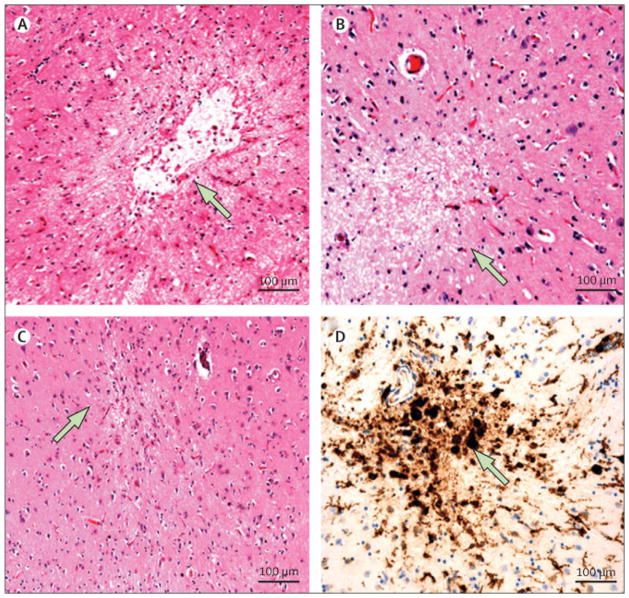
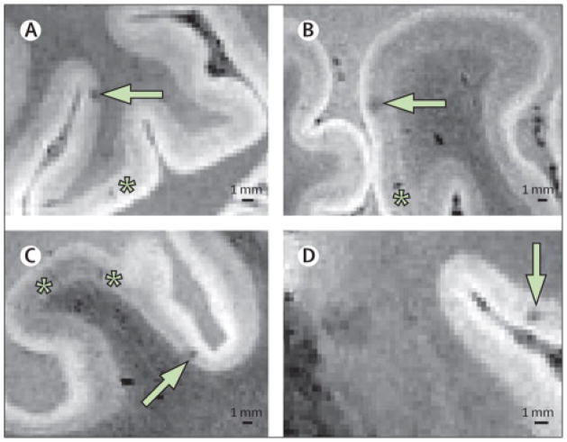
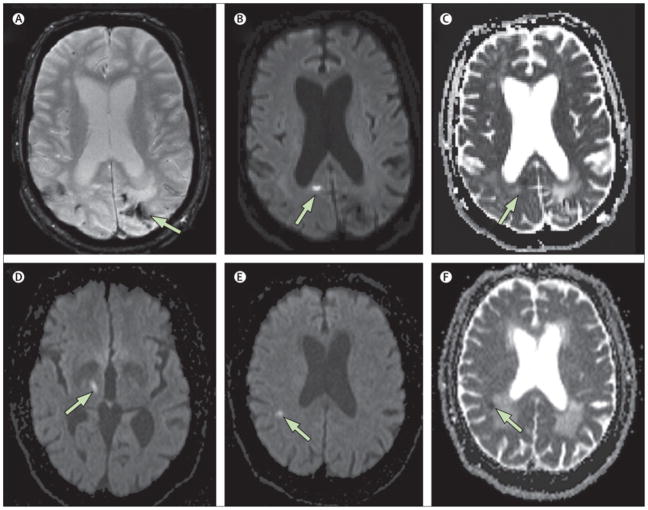
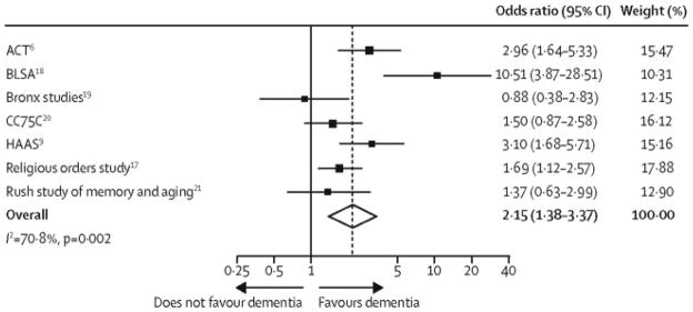
The association between small but still visible lacunar infarcts and cognitive decline has been established by population-based radiological and pathological studies. Microscopic examination of brain sections shows even smaller but substantially more numerous microinfarcts, the focus of this Review. These lesions often result from small vessel pathologies such as arteriolosclerosis or cerebral amyloid angiopathy. They typically go undetected in clinical-radiological correlation studies that rely on conventional structural MRI, although the largest acute microinfarcts can be detected by diffusion-weighted imaging. In view of their high numbers and widespread distribution, microinfarcts could directly disrupt important cognitive networks and thus account for some of the neurological dysfunction associated with lesions visible on conventional MRI such as lacunar infarcts and white matter hyperintensities. Standardised neuropathological assessment criteria and the development of non-invasive means of detection during life would be major steps towards understanding the causes and consequences of otherwise macroscopically invisible microinfarcts.


Conflict of interest statement

CONFLICT OF INTEREST STATEMENT

Dr. Smith reports no conflicts of interest. Dr. Schneider reports serving as a consultant to AVID Radiopharmaceuticals, and serving on advisory boards to Eli Lilly and Company and GE Healthcare. Dr. Wardlaw reports no conflicts of interest. Dr. Greenberg reports no conflicts of interest.

Figures

Figure 1
Figure 1. Microinfarct pathology
A. Cystic (cavitated) microinfarct (diameter: 600 μm) in the basal ganglia; original magnification 10x. B. Incomplete microinfarct without cavitation (diameter: 330 μm) in midfrontal cortex; original magnification 10x. C. Cortical microinfarct with linear scarring (“puckering”) from the middle temporal cortex (diameter 120 μm); original magnification 10x. D. Immunostatin for major histocompatibility complex II human leukocyte antigen DR3 (MHCII HLA-DR3) expression in activated microglia and macrophages in a microinfarct in the basal ganglia; original magnification 10X (diameter: 320 μm).
Figure 2
Figure 2. Intra-cortical Small Infarcts Detected on Post-Mortem High-Field Strength MRI
Examples of pathologically-verified intracortical microinfarcts (white arrows) imaged on post-mortem 7 tesla MRI (voxel size 0.3 mm3) of a 53 year old man who died of CADASIL. Asterisks indicate linear hypointensities pathologically proven to be perivascular spaces. The black bar indicates 1 mm. (Figure provided by E. Jouvent).
Figure 3
Figure 3. MRI Diffusion-Weighted Imaging of Small Acute Infarction (Sample)
Example of small acute areas of restricted diffusion detected incidentally on MRI. Top panels (A–C): A 70 year man with cerebral amyloid angiopathy who underwent MRI as part of a research study. Hemosiderin staining from prior hemorrhages is seen on the T2*-weighted gradient-recalled echo (GRE) sequence (A). Separate from these prior hemorrhages, an asymptomatic small hyperintensity is seen in the right occipital cortex on the diffusion-weighted image (DWI, panel B) with evidence of restricted diffusion on the apparent diffusion coefficient sequence (ADC, panel C), consist with an acute small cortical infarct. Bottom panels (D–F): A 67-year old man with an acute symptomatic lacunar infarct in the right thalamus (D, DWI sequence) also demonstrates an asymptomatic small 4.5 mm lesion in the right parietal white matter that is hyperintense on DWI (panel E) with restricted diffusion on ADC (panel F). An asymptomatic simultaneous small vessel infarct was suspected because no proximal source of embolism was identified and there was evidence of coexisting chronic cerebral small vessel disease (note confluent white matter lesions exhibiting increased diffusion in panel F).
Figure 4
Figure 4. Pooled Odds of Dementia in Persons with Microinfarcts (Unadjusted Analysis)
The unadjusted odds of dementia were more than doubled in the presence of microinfarcts (odds ratio 2.31, 95% confidence interval 1.40 to 3.82). Significant heterogeneity in the odds across studies (p=0.002) may be related to different methods for sampling for microinfarcts. See the legend to Table 1 for an explanation of the study acronyms.

References

    1. Matthews FE, Brayne C, Lowe J, McKeith I, Wharton SB, Ince P. Epidemiological pathology of dementia: attributable-risks at death in the Medical Research Council Cognitive Function and Ageing Study. PLoS Med. 2009;6:e1000180. - PMC - PubMed
    1. Vermeer SE, Longstreth WT, Jr, Koudstaal PJ. Silent brain infarcts: a systematic review. Lancet neurol. 2007;6:611–9. - PubMed
    1. Debette S, Markus HS. The clinical importance of white matter hyperintensities on brain magnetic resonance imaging: systematic review and meta-analysis. Bmj. 2010;341:c3666. - PMC - PubMed
    1. DerSimonian R, Laird N. Meta-analysis in clinical trials. Control Clin Trials. 1986;7:177–88. - PubMed
    1. Higgins JP, Thompson SG, Deeks JJ, Altman DG. Measuring inconsistency in meta-analyses. BMJ. 2003;327:557–60. - PMC - PubMed

Publication types

MeSH terms