. 2012 Jun;48(6):523-9.
doi: 10.1016/j.oraloncology.2012.01.010. Epub 2012 Feb 15.

Aberrant amplification of the crosstalk between canonical Wnt signaling and N-glycosylation gene DPAGT1 promotes oral cancer

Affiliations

Aberrant amplification of the crosstalk between canonical Wnt signaling and N-glycosylation gene DPAGT1 promotes oral cancer

Basem Jamal et al. Oral Oncol. 2012 Jun.

Abstract

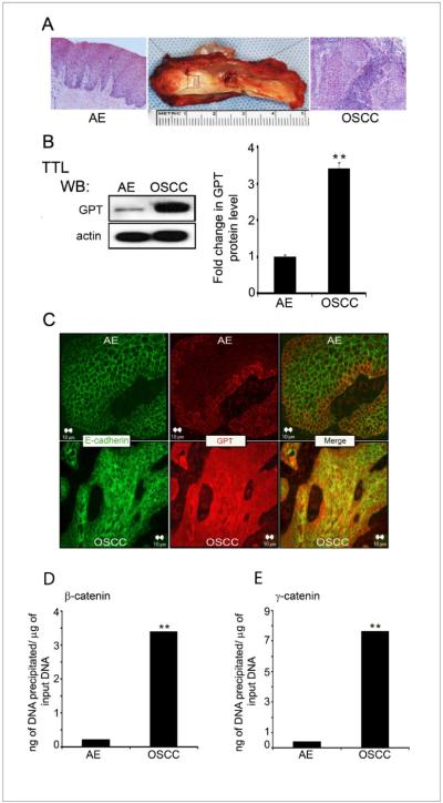
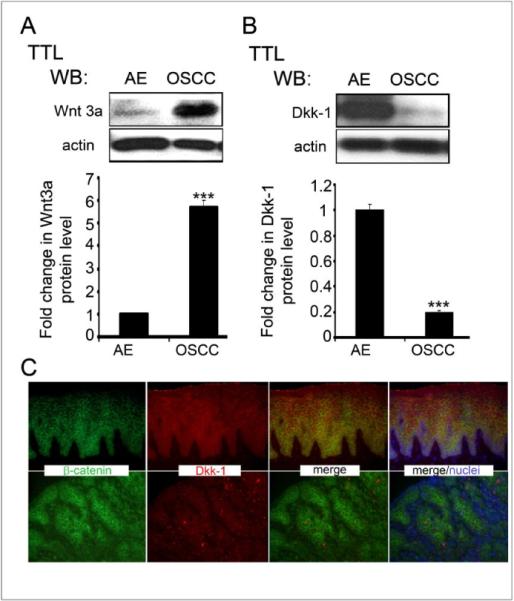
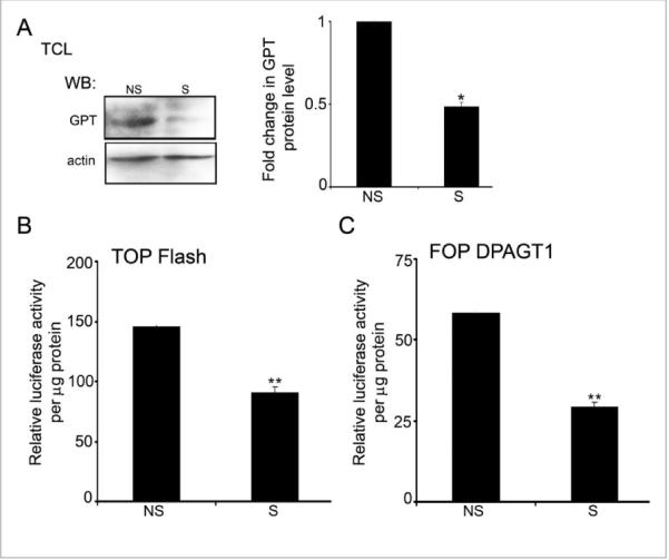
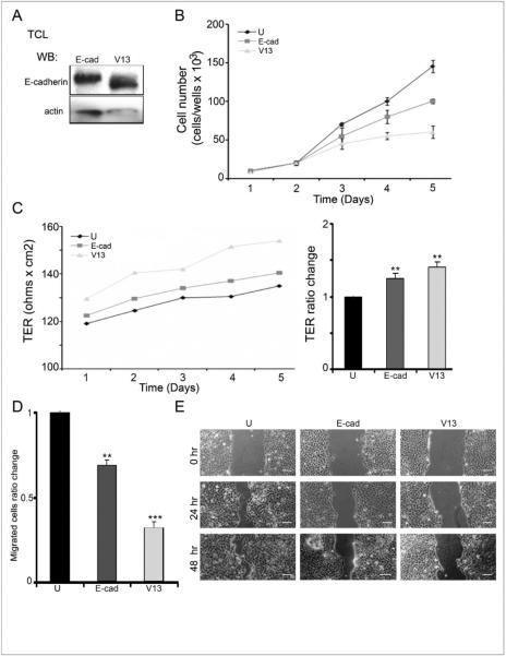
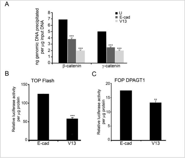
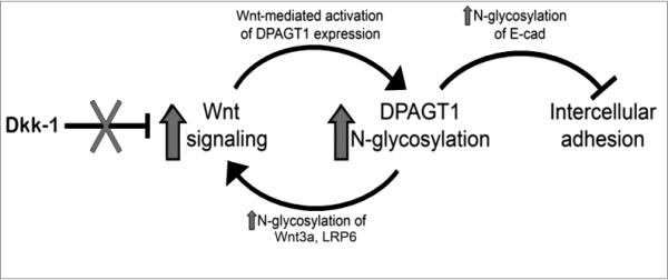
Oral cancer is one of the most aggressive epithelial malignancies, whose incidence is on the rise. Previous studies have shown that in a subset of human oral squamous cell carcinoma (OSCC) tumor specimens, overexpression of the DPAGT1 gene, encoding the dolichol-P-dependent N-acetylglucoseamine-1-phosphate transferase, a key regulator of the metabolic pathway of protein N-glycosylation, drives tumor cell discohesion by inhibiting E-cadherin adhesive function. Recently, we reported that DPAGT1 was a target of the canonical Wnt signaling pathway. Here, we link overexpression of DPAGT1 in human OSCC tumor specimens to aberrant activation of canonical Wnt signaling. We report dramatic increases in β- and γ-catenins at the DPAGT1 promoter and correlate them with reduced expression of a Wnt inhibitor, Dickkopf-1 (Dkk-1). Using human squamous carcinoma cell lines of the head and neck, we show that partial inhibition of DPAGT1 reduces canonical Wnt signaling, indicating that DPAGT1 and canonical Wnt signaling function in a positive feedback loop. We provide evidence that E-cadherin inhibits DPAGT1, canonical Wnt signaling and the OSCC cancer phenotype by depleting nuclear β- and γ-catenins, with hypoglycosylated E-cadherin being the most effective. This suggests that in human OSCC, extensive N-glycosylation of E-cadherin compromises its ability to inhibit canonical Wnt signaling and DPAGT1 expression. Our studies reveal a novel interplay between DPAGT1/N-glycosylation and canonical Wnt signaling and suggest that dysregulation of this crosstalk is a key mechanism underlying OSCC. They also suggest that partial inhibition of DPAGT1 may represent an effective way to restore normal interactions among these essential pathways in oral cancer.


Figures

Figure 1
Figure 1. Overexpression of DPAGT1 in OSCC is associated with increased promoter occupancy of β- and γ-catenins
(A) Composite resection specimen of OSCC and AE. (B) Western blot of GPT expression in AE and OSCC total tissue lysates (TTLs). Bargraph, Fold change in GPT levels after normalization to actin. (*p<0.05).(C) Immunofluorescence localization of E-cadherin and GPT in AE and OSCC. Size bar: 10 μm. (D) ChIP analysis of β-catenin at the DPAGT1 promoter in AE and OSCC(**p< 0.01). (E) ChIP analysis of γ-catenin at the DPAGT1 promoter in AE and OSCC (**p< 0.01). All results are from three independent experiments.
Figure 2
Figure 2. OSCC displays elevated tissue levels of β- and γ-catenins
(A) Western blot analysis of β-catenin and γ-catenin in AE and OSCC. Bargraph, Fold change in β-catenin and γ-catenin levels after normalization to actin. Results represent one of three experiments (*p<0.05). (B) Immunofluorescence localization of β- and γ-cateninsin AE and OSCC (arrows, insets).Size bar: 20 μm.
Figure 3
Figure 3. Upregulation of canonical Wnt in OSCC is associated with decreased Dkk-1 and elevated Wnt3a levels
(A) Western blot of Dkk-1 expression in AE and OSCC. Bargraph, Fold change in Dkk-1 levels after normalization to actin (***p< 0.001). (B) Western blot of Wnt3a expression in AE and OSCC. Bargraph, Fold change in Wnt3a levels in OSCC in comparison to AE after normalization to actin (**p< 0.01). (C) Immunofluorescence staining of Dkk-1 and b-catenin in AE and OSCC. Size bar: 50 μm. All results represent one of three independent experiments
Figure 4
Figure 4. Partial silencing of DPAGT1 inhibits canonical Wnt activity in A253 cells
(A) Western blot of GPT expression after partial silencing of DPAGT1with siRNA. Bargraph, Fold change of GPT abundance in silenced (S) and non-silenced (NS) cells after normalization to actin (*p< 0.05). (B) Luciferase reporter activity from the TOP Flash vector in S and NS cells (**p< 0.01). (C) Luciferase activity from the FOP DPAGT1 vector in S and NS cells(***p< 0.001). All results are from two independent experiments with each experiment repeated twice.
Figure 5
Figure 5. Hypoglycosylated E-cadherin variant, V13, interferes with CAL27 cell growth and migration and enhances intercellular adhesion
(A) Western blot of V13 and E-cad. (B) Growth curves of untransfected, E-cad- and V13-transfected cells (*p<0.05). (C) Transepithelial resistance (TER) in untransfected, E-cad- and V13-transfected cells (**p< 0.01).(D) Transwell migration assays of untransfected, E-cad- and V13-transfected cells (**p< 0.01 and ***p< 0.001). (E) Scratch- wound assays of untransfected, E-cad- and V13-transfected cells. Size bar: 50 μm.
Figure 6
Figure 6. Hypoglycosylated E-cadherin, V13, inhibits canonical Wnt activity and DPAGT1 transcription in CAL27 cells
(A) ChIP analyses using antibodies against β- and γ-catenins from E-cad and V13 cells (***p< 0.001). (B) Luciferase activity from the TOP Flash vector in E-cad and V13 cells (***p< 0.001). (C) Luciferase activity from the FOP DPAGT1 vector in E-cad and V13 cells (**p< 0.01). All results are from two independent experiments with each experiment repeated twice.
Figure 7
Figure 7. Interactions among canonical Wnt signaling, DPAGT1/N-glycosylation and E-cadherin in OSCC
Wnt signaling activates DPAGT1 and protein N-glycosylation, leading to extensive N-glycosylation of E-cadherin and weak intercellular adhesion. In OSCC, this positive feedback loop between Wnt signaling and DPAGT1is amplified by diminished expression of Dkk-1, acanonical Wnt inhibitor. Furthermore, extensive N-glycosylation of E-cadherin prevents it from depleting nuclear β- and γ-catenins, allowing the postitive feedback between Wnt and DPAGT1 to operate without controls.

Similar articles

Cited by

References

    1. Choi S, Myers JN. Molecular pathogenesis of oral squamous cell carcinoma: implications for therapy. J Dent Res. 2008;87:14–32. - PubMed
    1. Perez-Ordonez B, Beauchemin M, Jordan RC. Molecular biology of squamous cell carcinoma of the head and neck. J Clin Pathol. 2006;59:445–53. - PMC - PubMed
    1. Agrawal N, Frederick MJ, Pickering CR, Bettegowda C, Chang K, Li RJ, et al. Exome sequencing of head and neck squamous cell carcinoma reveals inactivating mutations in NOTCH1. Science. 2011;333:1154–7. - PMC - PubMed
    1. Stransky N, Egloff AM, Tward AD, Kostic AD, Cibulskis K, Sivachenko A, et al. The mutational landscape of head and neck squamous cell carcinoma. Science. 2011;333:1157–60. - PMC - PubMed
    1. Zuo J-H, Zhu W, Li M-Y, Li X-H, Yi H, Zeng G-Q, et al. Activation of EGFR Promotes Squamous Carcinoma SCC10A Cell Migration and Invasion via Inducins EMT-like PhenotypeCHange and MMP-9-Mediated Degradation of E-cadherin. J Cell Biochem. 2011;112:2508–17. - PubMed

Publication types

MeSH terms

Substances