. 2012 Apr;23(4):553-61.
doi: 10.1016/j.jvir.2011.12.017. Epub 2012 Feb 15.

Effects of Near-infrared Laser Irradiation of Biodegradable Microspheres Containing Hollow Gold Nanospheres and Paclitaxel Administered Intraarterially in a Rabbit Liver Tumor Model

Affiliations

Effects of Near-infrared Laser Irradiation of Biodegradable Microspheres Containing Hollow Gold Nanospheres and Paclitaxel Administered Intraarterially in a Rabbit Liver Tumor Model

Sanjay Gupta et al. J Vasc Interv Radiol. 2012 Apr.

Abstract

Purpose: To evaluate the effects of near-infrared (NIR) laser irradiation of microspheres (MS) containing hollow gold nanospheres (HAuNS) and paclitaxel (PTX) administered intraarterially in an animal model.

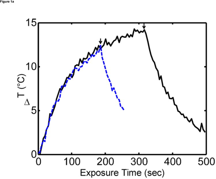
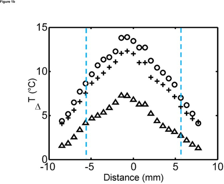
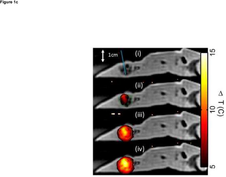
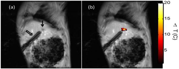
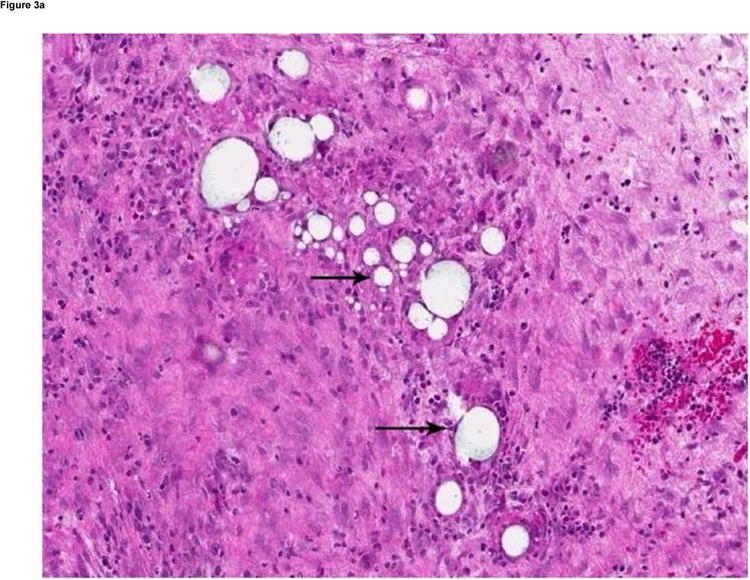
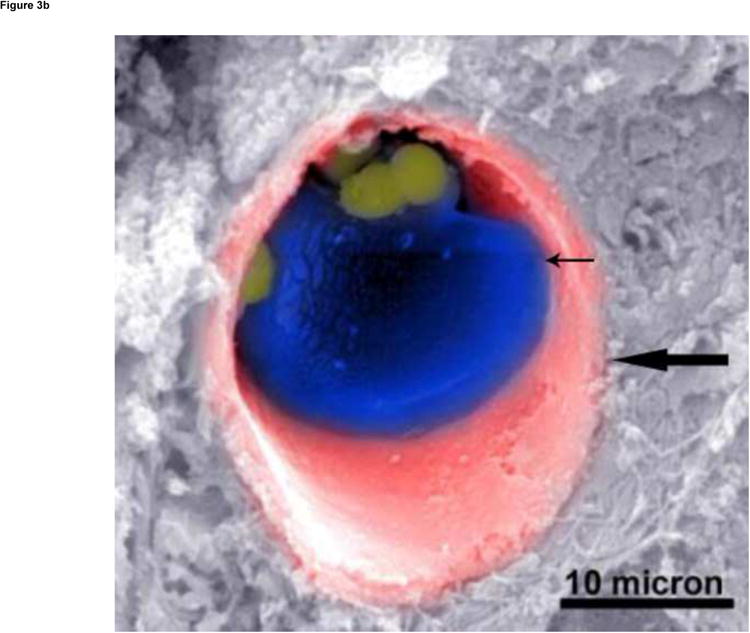
Materials and methods: For the ex vivo experiments, VX2 tumor-bearing rabbits underwent administration of MS-HAuNS or MS via the hepatic artery (HA). The animals were killed, the liver tumors were subjected to NIR irradiation, and temperature changes were estimated with magnetic resonance (MR) imaging. For the in vivo study, VX2 tumor-bearing rabbits were randomly assigned to three groups: MS-HAuNS-PTX-plus-NIR, MS-HAuNS-PTX, and saline-plus-NIR. Laser irradiation was delivered at 1 hour and at 3 days after administration of saline or MS-HAuNS-PTX via the HA. Animals were euthanized, and tumors were analyzed for necrosis and apoptosis. Plasma samples were collected from the MS-HAuNS-PTX-plus-NIR animals for PTX analysis.

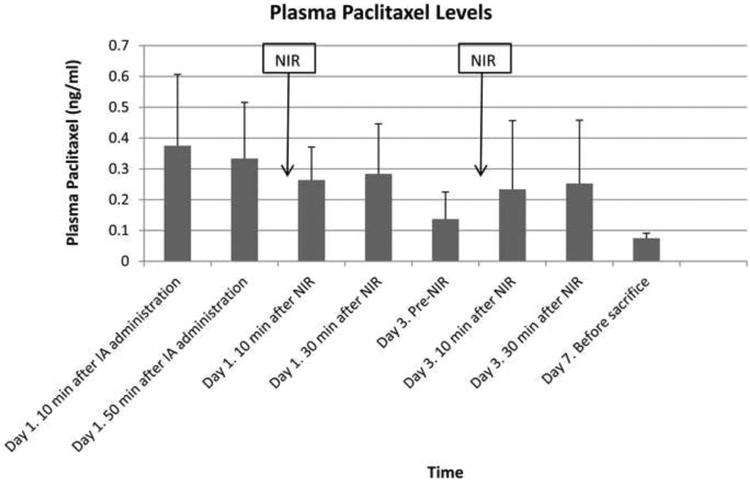
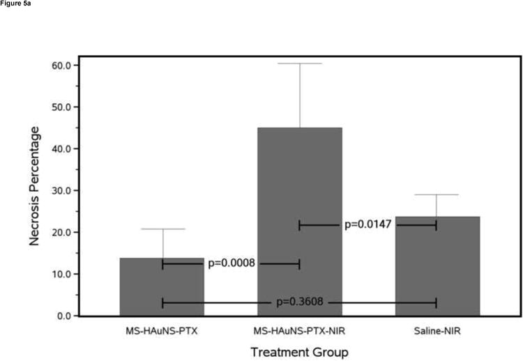
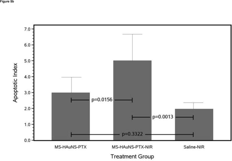
Results: Ex vivo experiments showed intratumoral heating in animals that received MS-HAuNS but no temperature change in animals that received MS. Animals treated with MS-HAuNS-PTX-plus-NIR showed a transient increase in plasma PTX levels after each NIR irradiation and significantly greater tumor necrosis than animals that received MS-HAuNS-PTX or saline-plus-NIR (44.9% vs 13.8% or 23.7%; P < .0001). The mean apoptotic index in the MS-HAuNS-PTX-plus-NIR group (5.01 ± 1.66) was significantly higher than the mean apoptotic index in the MS-HAuNS-PTX (2.99 ± 0.97) or saline-plus-NIR (1.96 ± 0.40) groups (P = .0013).

Conclusions: NIR laser irradiation after MS-HAuNS-PTX administration results in intratumoral heating and increases the efficacy of treatment. Further studies are required to evaluate the optimal laser settings to maximize therapeutic efficacy.


Figures

Figure 1
Figure 1
Ex vivo MR temperature imaging. (a) The estimated maximum change in temperature as measured by proton resonance frequency shift over time for a 180-sec (dashed) and a 300-sec (solid) exposure of 1.5 W at 808 nm. (b) Spatial profiles orthogonal to the laser fiber for 60 sec (triangle), 180 sec (+), and 300 sec (o) exposure times are shown versus the approximate tumor boundaries (dashed). (c) The approximate location of the laser fiber in the tumor (dashed line) is shown on the magnitude image of the fast chemical shift imaging acquisition (i). An overlay of the temperature change (5°C–15°C) as measured by proton resonance frequency for 60-sec (ii), 180-sec (iii), and 300-sec (iv) exposures of 1.5 W at 808 nm with an outline of the approximate tumor boundaries (green) shown for reference.
Figure 1
Figure 1
Ex vivo MR temperature imaging. (a) The estimated maximum change in temperature as measured by proton resonance frequency shift over time for a 180-sec (dashed) and a 300-sec (solid) exposure of 1.5 W at 808 nm. (b) Spatial profiles orthogonal to the laser fiber for 60 sec (triangle), 180 sec (+), and 300 sec (o) exposure times are shown versus the approximate tumor boundaries (dashed). (c) The approximate location of the laser fiber in the tumor (dashed line) is shown on the magnitude image of the fast chemical shift imaging acquisition (i). An overlay of the temperature change (5°C–15°C) as measured by proton resonance frequency for 60-sec (ii), 180-sec (iii), and 300-sec (iv) exposures of 1.5 W at 808 nm with an outline of the approximate tumor boundaries (green) shown for reference.
Figure 2
Figure 2
In vivo MR temperature imaging. Magnetic resonance (MR) temperature imaging of laser exposure (1.5 W for 180 sec) of VX2 in the liver of rabbits injected with MS-HAuNS-PTX. Sagittal MR image (a) shows the liver VX2 tumor (arrow) and the laser fiber inserted through a guide cannula (open arrow), whereas (b) shows the MR temperature imaging–estimated maximum temperature rise at the end of the exposure overlaid on the image.
Figure 3
Figure 3
(a) Hematoxylin and eosin–stained slide (180× magnification). MS are visualized as white spheres (black arrows) under light microscopy. (b) A scanning electron micrograph (3000× magnification) of a VX2 tumor shows the presence of MS (small arrow) in a tumor blood vessel (large arrow).
Figure 3
Figure 3
(a) Hematoxylin and eosin–stained slide (180× magnification). MS are visualized as white spheres (black arrows) under light microscopy. (b) A scanning electron micrograph (3000× magnification) of a VX2 tumor shows the presence of MS (small arrow) in a tumor blood vessel (large arrow).
Figure 4
Figure 4
Bar chart showing mean plasma PTX levels with 1 standard deviation error bars at different time points after IA administration of MS-HAuNS-PTX. The blood concentration of PTX was analyzed by using high-performance liquid chromatography.
Figure 5
Figure 5
Antitumor effects of various treatments on VX2 tumors grown in the liver of rabbits. (a) Bar chart of mean necrosis percentage for each treatment group with 1 standard deviation error bars and Tukey-Kramer adjusted pairwise P values. (b) Bar chart of mean apoptotic index for each treatment group with 1 standard deviation error bars and Tukey-Kramer adjusted pairwise P values.
Figure 5
Figure 5
Antitumor effects of various treatments on VX2 tumors grown in the liver of rabbits. (a) Bar chart of mean necrosis percentage for each treatment group with 1 standard deviation error bars and Tukey-Kramer adjusted pairwise P values. (b) Bar chart of mean apoptotic index for each treatment group with 1 standard deviation error bars and Tukey-Kramer adjusted pairwise P values.

References

    1. El-Sayed IH, Huang X, El-Sayed MA. Selective laser photo-thermal therapy of epithelial carcinoma using anti-EGFR antibody conjugated gold nanoparticles. Cancer Lett. 2006;239:129–135. - PubMed
    1. O'Neal DP, Hirsch LR, Halas NJ, Payne JD, West JL. Photo-thermal tumor ablation in mice using near infrared-absorbing nanoparticles. Cancer Lett. 2004;209:171–176. - PubMed
    1. Lu W, Xiong C, Zhang G, et al. Targeted photothermal ablation of murine melanomas with melanocyte-stimulating hormone analog-conjugated hollow gold nanospheres. Clin Cancer Res. 2009;15:876–886. - PMC - PubMed
    1. Melancon MP, Lu W, Yang Z, et al. In vitro and in vivo targeting of hollow gold nanoshells directed at epidermal growth factor receptor for photothermal ablation therapy. Mol Cancer Ther. 2008;7:1730–1739. - PMC - PubMed
    1. Bikram M, Gobin AM, Whitmire RE, West JL. Temperature-sensitive hydrogels with SiO2-Au nanoshells for controlled drug delivery. J Control Release. 2007;123:219–227. - PubMed