Review
. 2012 Mar;23(3):177-86.
doi: 10.1016/j.yebeh.2011.11.022. Epub 2012 Feb 16.

SCN1A mutations in Dravet syndrome: impact of interneuron dysfunction on neural networks and cognitive outcome

Affiliations
Review

SCN1A mutations in Dravet syndrome: impact of interneuron dysfunction on neural networks and cognitive outcome

Alex C Bender et al. Epilepsy Behav. 2012 Mar.

Abstract

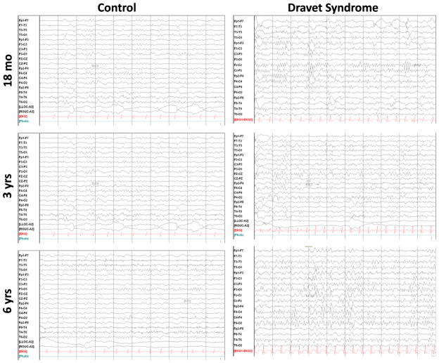
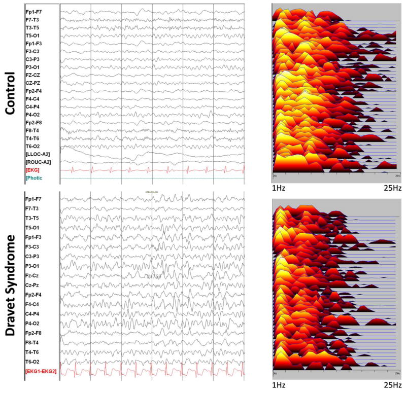
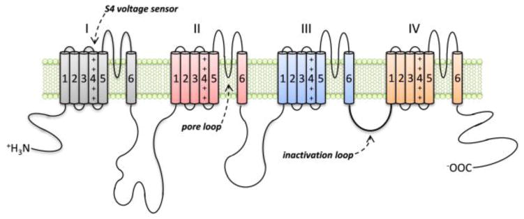
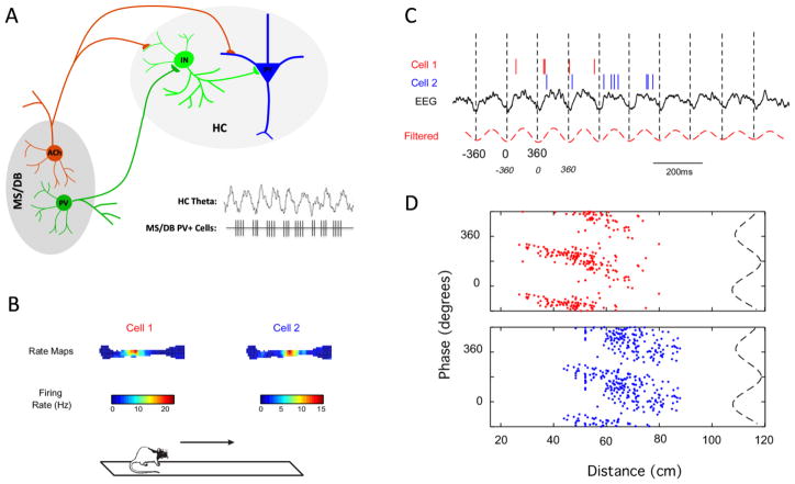
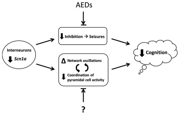
Dravet syndrome (DS) is a childhood disorder associated with loss-of-function mutations in SCN1A and is characterized by frequent seizures and severe cognitive impairment. Animal studies have revealed new insights into the mechanisms by which mutations in this gene, encoding the type I voltage-gated sodium channel (Na(v)1.1), may lead to seizure activity and cognitive dysfunction. In this review, we further consider the function of fast-spiking GABAergic neurons, one cell type particularly affected by these mutations, in the context of the temporal coordination of neural activity subserving cognitive functions. We hypothesize that disruptions in GABAergic firing may directly contribute to the poor cognitive outcomes in children with DS, and discuss the therapeutic implications of this possibility.


Figures

Figure 1
Figure 1
Examples of EEGs from age-matched healthy control and DS children. The EEGs are from six children. The children classified as controls had EEGs obtained because of possible seizures. All of these children had normal developmental assessments and neurological examinations. No discernible changes were noted between the child with DS and the control at 18 months of age. However, a progressive change is observed over the first 6 years. In both the 3 and 6 year-old children, the EEG had slower frequencies in the child with DS than the healthy controls, and this difference is most noticeable at 6 years of age.
Figure 2
Figure 2
EEG and power spectral analysis of a child with DS and a healthy control at 6 years of age. In the power spectral analysis on the right each line represents an electrode pair corresponding to the EEG on the left. Note that the spectral analysis of the child with DS was dominated by frequencies in the delta range whereas in the control, alpha range frequencies were prominent. Older children with DS typically have slower EEGs than age-matched controls.
Figure 3
Figure 3
The type I voltage-gated sodium channel (Nav1.1) alpha subunit. The alpha-subunit is composed of four repeated domains (I–IV) of six transmembrane segments each (S1–S6). The four pore loops between segments S5 and S6, which form the lining of the channel pore, confer selectivity of the channel for the sodium ion. The S4 segments act as the voltage sensors and activate opening of the channel in response to changes in membrane potential, and a loop on the cytosolic side of the membrane connecting domains III and IV is involved with channel inactivation. Both activation and inactivation of the channel are triggered by depolarization of the cell membrane and are characterized by fast kinetics. Opening of the channel produces an inward sodium conductance and rapid depolarization of the cell membrane, which are critical for initiating action potentials in neurons. Mutations in children with DS are often found in the key functional regions of the channel, particularly the S4 voltage sensors and S5–S6 pore loop regions.
Figure 4
Figure 4
The septo-hippocampal network and phase precession. A) The medial septum (MS) and diagonal band of Broca (DB), the hippocampal formation (HC) and the neural connections between these two brain regions through the fimbria fornix comprise the septo-hippocampal network. Cholinergic (Ach) and PV+ GABAergic neurons (PV) projecting from the MS/DB to the hippocampus are important for patterning and modulating theta oscillations. The population of PV+ projection neurons in the MS/DB fires in a bursting pattern, preceding the peak of each hippocampal theta cycle, and exerts a pacemaking role on hippocampal theta oscillations through the patterning of local hippocampal interneurons (IN). The significance of such temporal coordination of hippocampal network activity is well-illustrated by phase precession (B–D). B) Firing rate maps of two place cells recorded in the rodent hippocampus during performance on a linear track. C) While the rat crosses the linear track, place cells discharge at specific phases of the ongoing hippocampal EEG, shifting increasingly earlier in phase with each theta cycle. D) This phenomenon, termed phase precession, can be visualized by the phase-distance plots for each cell, with each theta cycle divided in 360º from trough to trough. Phase precession is believed to be important for spatial cognition by encoding the distance and event sequence as the animal traverses the corresponding place fields. Therefore, mutations in SCN1A that affect the firing of PV+ neurons in the septo-hippocampal network are likely to impair this important type of coordinated network activity.
Figure 5
Figure 5
A working model for possible contributions to cognitive impairment in DS. Loss-of-function mutations in SCN1A are found in children with DS. Animal studies have revealed that loss of function in Nav1.1, encoded by SCN1A, causes impaired firing of GABAergic interneurons relative to pyramidal cells. This is believed to cause an imbalance between excitation and inhibition, leading to seizures. The frequent and severe seizures observed in DS affect cognitive function. In addition, the functional deficit in GABAergic neurons likely alters the normal function of neural network activity known to be critical for cognitive functions. Examples may include alterations in theta and gamma oscillations, and loss of coordination of pyramidal cell ensembles involved in information processing. We hypothesize that these effects directly contribute to cognitive impairment independently of the contribution from seizures. Current antiepileptic drugs (AEDs) rebalance the relative amounts of excitation and inhibition in the brain to reduce the severity and frequency of seizures. However, it is unknown (“?”) whether treatments can ameliorate the alterations in network activity that are important for cognitive function. Future therapeutic strategies may benefit from consideration of these additional mechanisms that affect cognitive outcome.

Similar articles

Cited by

References

    1. Dravet C. Les epilepsies graves de l’enfant. La Vie medicale. 1978;8:543–8.
    1. Proposal for Revised Classification of Epilepsies and Epileptic Syndromes. Commission on Classification and Terminology of the ILAE. Epilepsia. 1989;30(4):389–99. - PubMed
    1. Hurst DL. Epidemiology of severe myoclonic epilepsy of infancy. Epilepsia. 1990;31(4):397–400. - PubMed
    1. Yakoub M, Dulac O, Jambaque I, Chiron C, Plouin P. Early diagnosis of severe myoclonic epilepsy in infancy. Brain & development. 1992;14(5):299. - PubMed
    1. Dravet C. The core Dravet syndrome phenotype. Epilepsia. 2011 Apr;52( Suppl 2):3–9. - PubMed

Publication types

MeSH terms