. 2012 Mar 9;45(5):629-41.
doi: 10.1016/j.molcel.2011.12.036. Epub 2012 Feb 16.

A mammalian autophagosome maturation mechanism mediated by TECPR1 and the Atg12-Atg5 conjugate

Affiliations

A mammalian autophagosome maturation mechanism mediated by TECPR1 and the Atg12-Atg5 conjugate

Dandan Chen et al. Mol Cell. .

Abstract

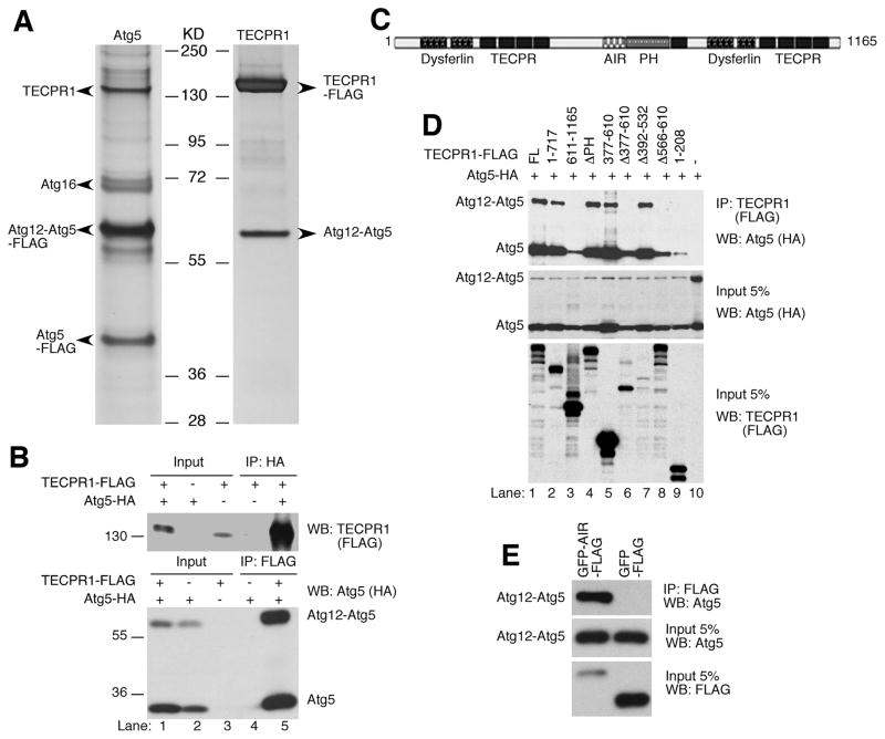
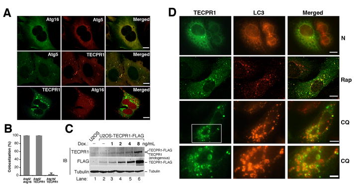
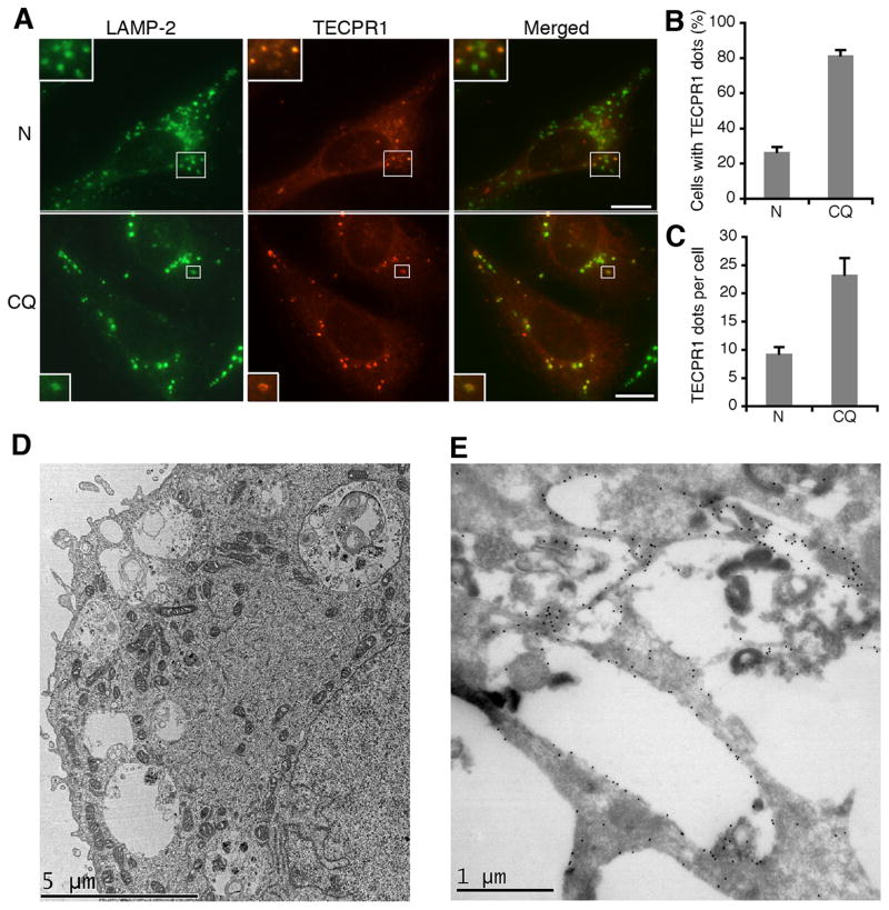
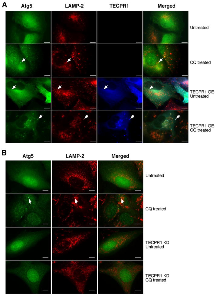
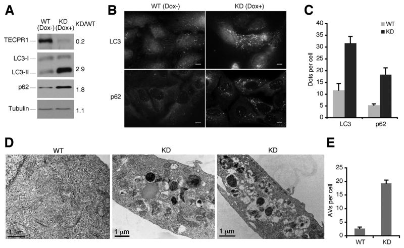
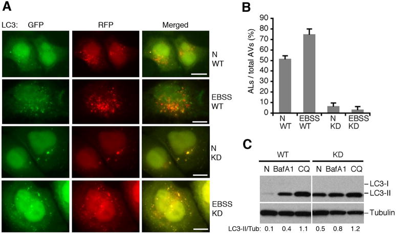
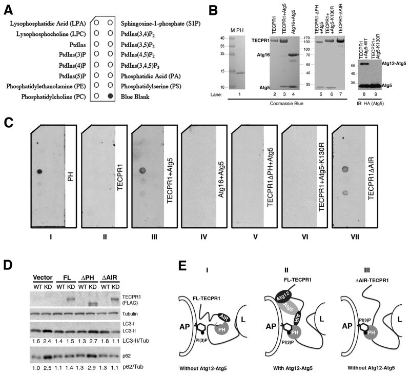
Autophagy is a major catabolic pathway in eukaryotes associated with a broad spectrum of human diseases. In autophagy, autophagosomes carrying cellular cargoes fuse with lysosomes for degradation. However, the molecular mechanism underlying autophagosome maturation is largely unknown. Here we report that TECPR1 binds to the Atg12-Atg5 conjugate and phosphatidylinositol 3-phosphate (PtdIns[3]P) to promote autophagosome-lysosome fusion. TECPR1 and Atg16 form mutually exclusive complexes with the Atg12-Atg5 conjugate, and TECPR1 binds PtdIns(3)P upon association with the Atg12-Atg5 conjugate. Strikingly, TECPR1 localizes to and recruits Atg5 to autolysosome membrane. Consequently, elimination of TECPR1 leads to accumulation of autophagosomes and blocks autophagic degradation of LC3-II and p62. Finally, autophagosome maturation marked by GFP-mRFP-LC3 is defective in TECPR1-deficient cells. Thus, we propose that the concerted interactions among TECPR1, Atg12-Atg5, and PtdIns(3)P provide the fusion specificity between autophagosomes and lysosomes and that the assembly of this complex initiates the autophagosome maturation process.


Conflict of interest statement

These authors have no conflict of interest.

Figures

Figure 1
Figure 1. TECPR1 interacts with the conjugated Atg12-Atg5
(A) Silver staining of the tandem affinity-purified Atg5 complexes or TECPR1 complexes in HEK293T cells. Proteins noted on the gel were identified by mass spectrometry. (B) Reciprocal co-immunoprecipitation of TECPR1 and Atg12-Atg5. HEK293T cells were transfected with TECPR1-FLAG and Atg5-HA. Whole cell lysate was immunoprecipitated with anti-FLAG or anti-HA agarose and analyzed by Western Blotting. (C) Domain structure of TECPR1. TECPR1 contains a pleckstrin homology (PH, aa611–717) domain, an Atg12-Atg5 interacting region (AIR, aa566–610), nine beta-propeller repeats (TECPR, aa209–240, 254–285, 301–332, 344–376, 729–756, 953–984, 1044–1075, 1087–1127), and two dysferlin domains (aa64–170, 816–922). (D) AA566–610 of TECPR1 is essential for Atg12-Atg5 binding. Atg5-HA was coexpressed with TECPR1-FLAG or different mutants as indicated. Whole cell lysate was immunoprecipitated with anti-FLAG agarose, followed by immunoblotting with anti-HA antibody. (E) The AIR domain of TECPR1 is sufficient for Atg12-Atg5 binding. Whole cell lysate was immunoprecipitated with anti-FLAG agarose. See also Figure S1.
Figure 2
Figure 2. TECPR1 localizes to late rather than early autophagic structures
(A) Co-staining of TECPR1-FLAG, Atg16-Myc or Atg5-HA in transfected U2OS cells. Scale bar (10 μm). (B) Quantification analysis of (A). Results are representative of three independent experiments and shown as means ± SD. (C) TECPR1-FLAG expression in U2OS cells was inducibly expressed by titrating doxycycline (DOX) as indicated for one day. Endogenously and ectopically expressed TECPR1 were detected by an anti-TECPR1 antibody. (D) Co-staining of TECPR1 and LC3 in U2OS cells. TECPR1-FLAG expression was induced at 2 ng/ml in U2OS cells that were either mock treated (N) or treated with rapamycin (Rap) (2 μM for four hours) or CQ (200 μM for two hours). Endogenous LC3 was detected using an anti-LC3 antibody. Scale bar (10 μm). The scale bar for the bottom panel of (D) is 5 μm. See also Figure S2.
Figure 3
Figure 3. TECPR1 localizes to the autolysosome membrane
(A) Co-staining of TECPR1 and LAMP-2 (upper, untreated; bottom, CQ treated (200 μM for two hours)) in U2OS cells expressing TECPR1-FLAG by 2 ng/ml doxycycline addition. An anti-LAMP-2 antibody was used to detect endogenous LAMP-2. Scale bar (10 μm). (B) Quantification of the percentage of cells with TECPR1 puncta described in (A). Results are representative of three independent experiments and presented as means ± SD. (C) Quantification of TECPR1 puncta per cell described in (A). Results are representative of three independent experiments and presented as means ± SD. (D) CQ treated (20 μM overnight) U2OS cells expressing EGFP-TECPR1 were processed and observed under the electron microscope. (E) CQ treated (20 μM overnight) U2OS cells expressing EGFP-TECPR1 were processed for cryo-immunolabeling. The anti-GFP primary antibody and gold conjugated secondary antibody were used to detect EGFP signals. See also Figure S3.
Figure 4
Figure 4. TECPR1 recruits Atg5 to autolysosomes
(A) TECPR1-FLAG was inducibly expressed by doxycycline (1 μg/ml) in U2OS cells co-expressing Atg5-EGFP. These cells were left untreated or treated with CQ (200 μM for two hours). Endogenous LAMP-2 was stained with a LAMP-2 antibody. Scale bar (10 μm). Representative Atg5-positive autolysosomes are marked by arrows. (B) In TECPR1 wild type and RNAi knockdown cells with or without CQ (200 μM for two hours) treatment, Atg5-EGFP and LAMP-2 staining were examined. Scale bar (10 μm). Representative Atg5-positive autolysosomes are marked by arrows. See also Figure S4.
Figure 5
Figure 5. Autophagosomes are accumulated in TECPR1 deficient cells
(A) Doxycycline-inducible TECPR1 RNAi depletion in U2OS-TR cells. In U2OS cells inducibly expressing shRNA against TECPR1, endogenous TECPR1, LC3, p62 and Tubulin were detected in the presence or absence of doxycycline (1 μg/ml) for 5 days. (B) Endogenous LC3 or p62 was stained using anti-LC3 or anti-p62 antibody. Scale bar (10 μm). (C) Quantification result of at least 200 cells from three independent experiments described in (B). Data are presented as means ± SD. (D) Control or TECPR1 depleted U2OS cells were analyzed under transmission electron microscope. (E) Quantification result of at least 100 cells from three independent experiments described in (D). Data are presented as means ± SD. See also Figure S5.
Figure 6
Figure 6. TECPR1 is required for autophagosome maturation
(A) GFP-mRFP-LC3 was expressed and detected 48 hours after transfection in control cells or TECPR1 depleted U2OS cells treated with EBSS medium for one hour or mock treated. Scale bar (10 μm). (B) Quantification result of at least 200 cells from three independent experiments described in (A). Data are presented as means ± SD. AL, autolysosomes; AV, autophagic vacuoles. (C) The autophagic flux was examined in control and TECPR1 depleted U2OS cells mock treated (N), BafA1 or CQ treated. The intensity of LC3II/Tubulin was calculated as shown. See also Figure S6.
Figure 7
Figure 7. Binding of PtdIns(3)P by TECPR1 requires Atg12-Atg5 association
(A) Schematic of a PIP Strip spotted with eight phosphoinositides and other bioactive lipids. (B) Purification of recombinant PH domain (lane 1), full-length TECPR1 (lane 2), Atg12-Atg5-TECPR1 complex (lane 3), Atg12-Atg5-Atg16 complex (lane 4), Atg12-Atg5-TECPR1 (PH domain deleted) (lane 5), TECPR1-Atg5K130R (lane 6) and Atg12-Atg5-TECPR1 (AIR domain deleted) (lane 7) in transfected HEK293T cells. Purified recombinant Atg12-Atg5-TECPR1 complex (lane 8) and TECPR1-Atg5K130R complex (lane 9) were probed with anti-Atg5 (HA) antibody for Western Blotting. (C) PIP Strips were incubated with recombinant proteins (1 μg/ml) as indicated for overnight at 4°C and probed with anti-FLAG antibody. (D) Complementation of TECPR1 with the indicated shRNA-resistant TECPR1 expression plasmids in TECPR1 depleted U2OS-TR cells. The inducible U2OS cells were cultured in the presence or absence of doxycycline (1 μg/ml) for 5 days. Expression of TECPR1 wild-type and mutants was detected with the FLAG antibody. The intensities of LC3II/Tubulin and p62/Tubulin were calculated as shown. (E) Proposed models of the role of TECPR1 in autophagosome maturation through its association with Atg12-Atg5 and PtdIns(3)P. Panel I, full-length TECPR1 in the absence of the Atg12-Atg5 conjugate. Panel II, full-length TECPR1 in the presence of the Atg12-Atg5 conjugate. Panel III, AIR-deleted TECPR1 in the absence of the Atg12-Atg5 conjugate. AP, autophagosome; L, lysosome. See also Figure S7.

References

    1. Abeliovich H, Darsow T, Emr SD. Cytoplasm to vacuole trafficking of aminopeptidase I requires a t-SNARE-Sec1p complex composed of Tlg2p and Vps45p. Embo J. 1999;18:6005–6016. - PMC - PubMed
    1. Backer JM. The regulation and function of Class III PI3Ks: novel roles for Vps34. Biochem J. 2008;410:1–17. - PubMed
    1. Baerga R, Zhang Y, Chen PH, Goldman S, Jin S. Targeted deletion of autophagy-related 5 (atg5) impairs adipogenesis in a cellular model and in mice. Autophagy. 2009;5:1118–1130. - PMC - PubMed
    1. Behrends C, Sowa ME, Gygi SP, Harper JW. Network organization of the human autophagy system. Nature. 2010;466:68–76. - PMC - PubMed
    1. Boonen M, van Meel E, Oorschot V, Klumperman J, Kornfeld S. Vacuolization of mucolipidosis type II mouse exocrine gland cells represents accumulation of autolysosomes. Mol Biol Cell. 2011;22:1135–1147. - PMC - PubMed

Publication types

MeSH terms

Substances